EJCRIM 2023 CiteScore
| 2.1 = | 1.762 Cit. to date |
| 842 Docs. to date |
Last updated on 05 May, 2024
Updated monthly
Updated monthly
Powered by

|
Views: 58
HTML: 3
PDF: 94
|
Chylothorax is the accumulation of lymphatic fluid (chyle) within the pleural space. There are multiple causes, including traumatic and non-traumatic mechanisms. Trauma can cause disruption of the thoracic duct either by direct damage or indirect crushing or avulsion mechanisms. Non-traumatic causes include infections, inflammatory processes, malignancies, and iatrogenic injury (during surgery or central venous access). The traditional management of traumatic chylothorax has been either a conservative approach, including complete nil per os, or a low-fat diet with medium-chain triglyceride supplementation with the administration of somatostatin or its analog, octreotide, versus a surgical approach consisting of thoracic duct ligation. Recently a less invasive approach via thoracic duct embolization has gained popularity. There have been a few reports of the successful use of an alpha 1-adrenergic agonist (midodrine) as an adjunct in the conservative approach. We describe the utility of midodrine in three cases of chylothorax and propose a management algorithm.
|
Views: 35
HTML: 3
PDF: 16
|
Atrial myxoma is a rare primary tumour of the heart that typically arises from the left atrium. Patients typically present with obstructive symptoms such as dyspnoea, but constitutional and embolic symptoms can be seen as well. Gastrointestinal symptoms in the absence of embolisation are rarely reported in the literature. Our case presents a 55-year-old female who was found to have a large left atrial myxoma after presenting with gastrointestinal symptoms, which resolved upon resection of the tumour. This case illustrates that atrial myxomas can have an atypical presentation with gastrointestinal symptoms, which could be related to inflammation of gastric mucosa from interleukin-6 produced by the tumour cells. Careful history-taking followed by early detection and prompt treatment is important as atrial myxomas can lead to potentially devastating complications.
|
Views: 41
HTML: 4
PDF: 13
|
Background: Heterotopic splenic tissue can occur following splenectomy and is typically asymptomatic, often discovered incidentally during imaging for other conditions. This benign condition may mimic malignant processes, posing diagnostic challenges especially in patients with a history of cancer or concurrent malignancy.
Case description: We report the case of a 60-year-old male with a history of well-controlled hypertension and a splenectomy following a traumatic injury at age 7. The patient underwent routine screening which revealed elevated prostate-specific antigen (PSA) levels. Subsequent magnetic resonance imaging (MRI) identified suspicious lesions in the prostate and a left lower quadrant mass. Prostate biopsy confirmed an adenocarcinoma with a Gleason score of 6, while biopsy of the abdominal mass revealed heterotopic splenic tissue. The management strategy included active surveillance for prostate cancer, considering the tumour’s low aggressiveness and the benign nature of the splenic tissue.
Conclusions: This case highlights the importance of considering heterotopic splenic tissue in differential diagnosis when evaluating abdominal masses in patients with a history of splenectomy. Accurate diagnosis through careful imaging and biopsy is crucial to avoid misdiagnosis and unnecessary treatments, particularly in patients with concurrent malignancies.

|
Views: 62
HTML: 5
PDF: 21
|
Ischaemic colitis is responsible for more than half of the presentations of gastrointestinal ischaemia and develops due to an interruption of intestinal blood flow. Risk factors include increasing age and conditions associated with decreased perfusion. Infrequently, ischaemic colitis may develop in young females prescribed oral contraceptives. Here, we present a case of ischaemic colitis secondary to oral contraceptives that resolved with medication discontinuation.
|
Views: 128
HTML: 121
PDF: 35
|
Background: While the use of immunotherapy has revolutionised the treatment of various cancers, it is often associated with a myriad of immune-related adverse effects.
Case Presentation: In this article, we report a rare case of durvalumab-induced triple-M syndrome in a 69-year-old woman with stage III lung adenocarcinoma. She was admitted with profound generalised muscle weakness, myalgia, and exertional breathlessness, about a week into her second cycle of durvalumab, an immune checkpoint inhibitor. She had clinicopathological features of myositis, myasthenia and myocarditis with acute onset symptomatic tri-fascicular block on electrocardiogram, requiring urgent cardiology intervention. Durvalumab was discontinued and she was treated with a combination of high-dose steroids and intravenous immunoglobulin after which she had clinical and biochemical improvement, albeit with residual muscle weakness.
Conclusion: Myocarditis-myositis-myasthenia complex is a rare side effect of immunotherapy which has been reported in other immune checkpoint inhibitors, but less so with durvalumab. We report this clinical case to raise awareness of this rare and potentially life-threatening adverse effect of this agent.

|
Views: 67
HTML: 12
PDF: 46
|
Subarachnoid haemorrhage (SAH) is a rare yet consequential medical emergency that may mimic an acute myocardial infarction (MI). SAH causes enhanced sympathetic activity, culminating in the development of neurogenic stunned myocardium (NSM), which presents as ST-segment deviations, prolonged QT intervals, T-wave inversions or Q-waves. Reperfusion therapy is contraindicated for SAH because of an increased risk of bleeding and death. Therefore, a prompt diagnosis is crucial. Here, we report a unique case of massive SAH presenting as diffuse ST-segment deviation simulating an acute MI. Our patient was brought to the emergency department after a cardiac arrest and died on day 2 of admission.
|
Views: 34
HTML: 2
PDF: 12
|
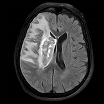
Background: Aphasia is a common neurocognitive disorder caused by impaired speech and language, with stroke being the most frequent cause. The neuroanatomical mechanism underlying this condition is not yet fully understood.
Case description: This case describes a 74-year-old Caucasian woman admitted with a clinical picture of right total anterior circulation infarct (TACI) and aphasia, scoring 17 on the National Institutes of Health Stroke Scale. Neuroimaging showed a large cortico-subcortical frontotemporoparietal and insular infarct involving the basal ganglia of the right hemisphere and bilateral focal atherosclerotic stenosis on the M1 segment of the middle cerebral artery. There was no left hemispheric lesion or abnormal electric activity on the electroencephalogram. A formal evaluation was compatible with transcortical motor aphasia. The aetiological study revealed atrial fibrillation, and the case was admitted as an ischaemic stroke of undetermined aetiology with two possible causes – intracranial atherosclerotic stenosis or atrial fibrillation.
Conclusion: Our patient fulfilled all the formal criteria for crossed aphasia in dextral (CAD): aphasia, a lesion in the right hemisphere coupled with the structural integrity of the left hemisphere, an established preference for right-hand use without a familial history of left-handedness individuals, and an absence of brain damage in childhood. Our patient’s case adds to the evidence that deep structures – alone or in combination with cortical structures – are primarily affected in CAD.

|
Views: 50
HTML: 6
PDF: 19
|
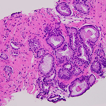
Background: Hairy cell leukaemia (HCL) is an uncommon, indolent, B-cell, lymphoproliferative disorder typically involving peripheral blood, spleen and bone marrow. It is commonly presenting with pancytopenia, monocytopenia and massive splenomegaly, while accounting for 2% of lymphoid leukaemias. Cases of extranodal lesions caused by HCL are rare, although these have been reported. Here, we report a case of HCL presenting as a paravertebral mass without systemic involvement.
Case description: A 58-year-old man was admitted to our hospital due to progressive difficulty walking for a month, without any other symptoms. Blood examination noted mild anaemia with Hb=12.6 g/dl and mild thrombocytopenia of 140,000/ul. Magnetic resonance imaging (MRI) and computed tomography (CT) imaging demonstrated a T6 posterior paravertebral mass lesion, extending into the spinal canal with metastatic bone lesions along the thoracic and lumbar spine. Further imaging study with CT indicated mild splenomegaly (13.4 cm) and an enlarged abdominal lymph node (3.5 cm) near celiac trifurcation.
Conclusion: A core-needle biopsy from the paravertebral mass was performed. Results showed small-sized cells with round or oval nuclei, and pale cytoplasm with immunophenotype: B-cell origination with CD20+, Cyclin D1+, DBA.44+, Annexin+ and BRAF+, indicative of HCL.
|
Views: 45
HTML: 4
PDF: 15
|
We present the case of a 63-year-old female diagnosed with atypical SSc in the setting of acute SRC. She was undergoing work-up for progressive dyspnoea in the outpatient setting when she was found to have newly diagnosed restrictive lung pathology and worsening renal function, thus prompting acute hospital admission. Given multisystem involvement of the pulmonary and renal systems, the differential diagnosis included autoimmune and connective tissue disorders. Although serologies were non-specific, renal biopsy confirmed scleroderma renal disease, and she was started on treatment with captopril. This case highlights the importance of clinical judgment and timely diagnosis, even when laboratory data might indicate otherwise.

|
Views: 114
HTML: 23
PDF: 55
|
Legionella pneumophila is a bacterium that usually causes pulmonary disease but can rarely present with extrapulmonary manifestations, such as rhabdomyolysis. This is a case of Legionella infection with significant rhabdomyolysis but a lack of acute kidney injury.
A 38-year-old male with a history of epilepsy presented to the emergency department after a seizure episode with confusion, fever, emesis and bruises. He also complained of a productive cough and scant haemoptysis for the past two months. Chest X-ray showed retrocardiac and left upper lobe opacities; urine was positive for Legionella antigen and myoglobinuria. Creatinine phosphokinase was 242,488 U/l and creatinine was 0.5 mg/dl. The patient was managed with oxygen therapy, aggressive IV hydration and IV azithromycin, and later IV levofloxacin until his symptoms resolved.
Rhabdomyolysis may be a sign of Legionella infection. Rapid testing of Legionella antigen, especially in populations at risk, may be crucial for timely diagnosis and treatment. Kidney function may be preserved in the early stages of disease, but early treatment with antibiotics and aggressive hydration are an effective way to prevent deterioration in kidney function.

|
Views: 81
HTML: 11
PDF: 39
|
Background: Acute pancreatitis is a common cause of hospitalisation characterised by inflammation of the pancreas. While mechanical, toxic and iatrogenic factors typically cause it, post-oesophagogastroduodenoscopy (EGD) pancreatitis is extremely rare. This report examines a case of acute pancreatitis following EGD, aiming to highlight this rare but significant complication.
Case description: A 46-year-old woman with a history of breast cancer, anxiety, vitamin D deficiency and gastro-oesophageal reflux disease underwent an EGD, which revealed and led to the removal of duodenal polyps. Six hours post-procedure, she presented with severe abdominal pain radiating to her back, accompanied by nausea. Laboratory results indicated elevated lipase levels, and a computed tomography (CT) scan confirmed acute pancreatitis. The patient was managed with aggressive fluid resuscitation, bowel rest and pain management, leading to an improvement in her condition and subsequent discharge. We believe that the pancreatitis was likely caused by the use of cautery during the endoscopic mucosal resection of duodenal polyps.
Conclusion: This case underscores the need for clinicians to recognise acute pancreatitis as a potential complication of EGD, especially in the absence of other common risk factors.
|
Views: 229
HTML: 31
PDF: 41
|
Gastric carcinoid is a rare type of gastric malignancy accounting for around 7% of all gastrointestinal neuroendocrine tumours (NETs). While most gastric NETs (gNETs) are readily visible through direct visualisation by upper endoscopy, around 25% of gastric carcinoids are invisible because they are located in the submucosal gastric regions of the body and fundus. gNETs located in the intra-mucosal areas can be identified by gastric mapping; this can be done by taking random gastric biopsies from the antrum, body and fundus.
We report a case of a well-differentiated gastric NET type 1 with atrophic gastritis diagnosed on upper endoscopy and pathological immunohistochemistry staining.

|
Views: 54
HTML: 1
PDF: 36
|
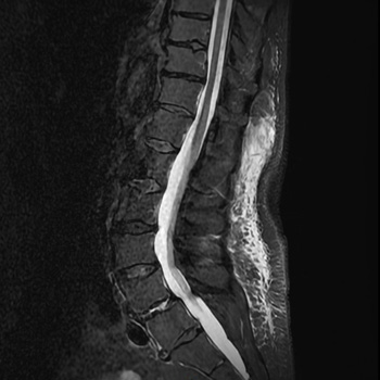
Background: Arteriovenous malformations (AVMs) are abnormal direct connections between arterial and venous systems, without an interposed capillary bed. This permits high-flow arteriovenous shunting, which precipitates structural changes in the afferent and efferent vessels, namely arterial smooth muscle hyperplasia and thinning of venous walls. Patients with intracranial AVMs typically present with a haemorrhage, headache or seizure. Treatment is either via medical management aimed at control of seizures, headache and blood pressure, or interventional via surgical, radiation or radiologically guided embolisation.
Case description: We report the case of a woman in her early 40s presenting with a tonic-clonic seizure against a background of a 31-year history of migraine and an 18-month history of tremors in her right arm. The clinical examination was remarkable for an extremely loud cranial bruit and a right homonymous hemianopia. Imaging diagnosed an 8 cm Martin-Spetzler grade V intracranial arteriovenous malformation in her left parietal lobe, which was deemed unsuitable for operative or radiotherapy-based intervention.
Conclusion: The patient was managed through observation and relatively good control of her breakthrough seizures was achieved through the addition of brivaracetam to her lamotrigine and carbamazepine-based therapy, six years after her initial presentation.

|
Views: 98
HTML: 4
PDF: 26
|
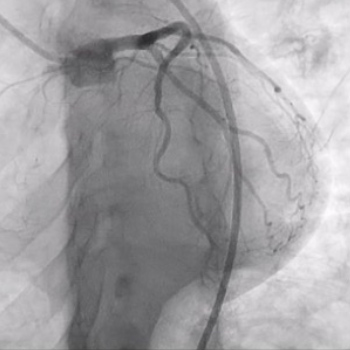
Introduction: Aortic pseudoaneurysms are a type of contained rupture where most of the aortic wall is breached, leaving only a thin rim of the remaining wall or adventitia to hold the blood. This condition carries a high risk of rupture and potentially fatal complications. Typically, patients present with chest pain; haemoptysis can also occur, though rarely.
Case description: A 64-year-old male who presented with two episodes of haemoptysis, with no history of cardiovascular surgery or trauma. A chest computerized tomography (CT) followed by an aortogram revealed a thoracic aortic pseudoaneurysm and the patient underwent surgical aortic repair without any complications. This case underscores the rare presentation of thoracic aortic pseudoaneurysm.
Discussion: Haemoptysis is a rare manifestation of thoracic aorta pseudoaneurysm and can be a warning sign of impending rupture. Haemoptysis may occur due to formation of aortopulmonary fistula or direct erosion of pseudoaneurysm into lung parenchyma.
Conclusion: It is imperative for clinicians to recognise such manifestations early for prompt diagnosis and prevention of complications.

|
Views: 53
HTML: 7
PDF: 15
|
Gastric intramural haematoma is a very infrequent condition. It can occur due to clotted gastric haemorrhage as a result of peptic ulcer disease, or following trauma, oral anticoagulant therapy and bleeding disorders. It is usually suspected with the symptoms of gastrointestinal haemorrhage such as haematemesis, melena and haematochezia, and detected by endoscopy. In rare cases, the patient is asymptomatic or presents with non-related symptoms and can diagnosed by computerised tomography. In this case, we report the detection of a gastric intramural haematoma during abdominal ultrasonography in a hypotensive patient who was admitted to the emergency department after sliding and falling from a height.

|
Views: 580
HTML: 31
PDF: 156
|
Haemorrhagic pleural effusion can be a challenging diagnosis that requires a thorough investigation and sometimes a multidisciplinary team of physicians to reach the underlying aetiology. Causes can include pulmonary malignancy, pulmonary infections, connective tissue diseases, asbestos associated, intra-abdominal conditions such as pancreatitis and ovarian tumours, cardiovascular disorders such as ruptured aneurysms and pulmonary infarction, as well as other miscellaneous causes. One such cause is endometriosis in the thoracic cavity. Endometriosis is a chronic illness associated with the occurrence of endometrial tissue outside the endometrium. Insertion of endometrial tissue in the thoracic cavity is rare, with only a few cases described. This case report gives detail of a 30-year-old nulligravida suspected of having thoracic endometriosis following a history of catamenial dyspnoea and associated pleural effusion. The diagnosis was confirmed through the histopathological study of tissue obtained via thoracoscopic surgery. Excision of the endometrial tissue was done, and the patient then continued medical treatment with progestins and gonadotrophin-releasing hormone (GnRH) agonists. Following therapy, the index patient was asymptomatic. A multidisciplinary approach is often needed in the diagnosis and management of thoracic endometriosis, involving both medical and surgical specialities. Minimally invasive surgery is the gold standard of diagnosis, allowing for direct visualisation of implants and nodules and should be followed by medical treatment to reduce the risk of recurrence. Medical therapy alone is associated with higher rates of recurrence. Physicians must have a high degree of suspicion as thoracic endometriosis is a disease that can often be missed.

|
Views: 62
HTML: 15
PDF: 46
|
Tumour-to-tumour metastasis (TTM) is a rare phenomenon that clinicians should be aware of when evaluating patients with a history of prostate cancer. We present the diagnosis and management of an 80-year-old former smoker with high-risk prostate cancer, who developed a lung nodule consistent with TTM. The patient had concurrent primary lung adenocarcinoma and metastatic prostate cancer, making this a unique case of dual primary and metastatic malignancies. The complexity of this case highlights the need for comprehensive evaluation and interdisciplinary management in patients with multiple malignancies. The literature review reveals that these are extremely rare occurrences, with most cases involving metastasis to the second primary tumour. Despite the challenges in diagnosing preoperatively, it is important to consider TTM as a possibility in patients with prostate cancer who present with a lung nodule. This report presents one of the few documented cases of TTM. It also reviews relevant cases in the literature and discusses the current situation in relation to established criteria for classifying combination tumours.

|
Views: 76
HTML: 9
PDF: 75
|
Takayasu arteritis (TA) primarily causes ischaemic nephrosclerosis but can occasionally be associated with glomerulopathy. We report a case of a female in her twenties with PLA2-negative, THSD7A-positive membranous nephropathy (MN) refractory to rituximab, who presented with neck pain and new-onset hypertension. Blood work showed elevated inflammatory markers. Imaging of the head and neck revealed focal dilation and irregularity of the vertebral arteries, consistent with TA. The patient was started on treatment with steroids, followed by mycophenolate mofetil, which led to the resolution of symptoms and nephrotic syndrome. This case highlights an uncommon sequence of events, with MN presenting before TA, underscoring the need to consider TA in differentials for patients with MN. Notably, this is the first reported case in a young female, emphasising the need for further understanding of TA-associated glomerular diseases. Additionally, the presence of THSD7A in MN, despite negative malignancy workup, is also noteworthy.
| 2.1 = | 1.762 Cit. to date |
| 842 Docs. to date |


Publisher
Official Journal of the
European Federation of Internal Medicine
www.efim.org
Publisher: SMC media Srl
Via Giovenale, 7 - 20136 Milan - Italy
P.IVA 07626490960
info@ejcrim.com
www.ejcrim.com - ISSN: 2284-2594 - © EFIM 2014-2024, Published by SMC Media srl, Italy - Privacy policy

