
EJCRIM 2023 CiteScore
| 2.1 = | 1.730 Cit. to date |
| 842 Docs. to date |
Last updated on 05 March, 2024
Updated monthly
Updated monthly
Powered by

|
Views: 129
HTML: 23
PDF: 177
|
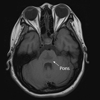
Osmotic demyelination syndrome (ODS) is a disorder characterised by the widespread development of demyelination in both pontine and extrapontine regions. It has been recognised as a complication arising from the rapid correction of hyponatraemia. This study presents the case of a 20-year-old Thai female patient at 10 weeks gestation, exhibiting an initial presentation of catatonia – an uncommon manifestation of ODS. The patient developed symptoms following the rapid correction of hyponatraemia in the context of hyperemesis gravidarum. Magnetic resonance imaging (MRI) of the brain revealed a trident or bat-wing-shaped pattern in T2-weighted and fluid-attenuated inversion recovery (FLAIR) sequences at the central pons. The patient underwent five cycles of plasmapheresis and received rehabilitation, leading to clinical improvement.
|
Views: 80
HTML: 5
PDF: 57
|
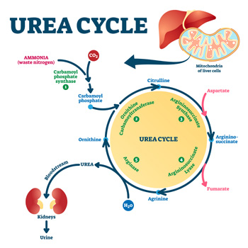
Background: Hyperargininemia is a rare inherited metabolic disorder of the urea cycle with an autosomal recessive transmission. It occurs due to a deficiency of the enzyme arginase I and causes progressive neurological damage. Very few cases are diagnosed in adulthood, with the majority being diagnosed before the age of 4. Currently, this condition is diagnosed by a mass spectrometry technique in neonatal screening, which has been implemented in Portugal since 2007; births before that were not screened for this entity.
Case description: We present a case of a 23-year-old woman referred to the internal medicine and neurology departments with a history of two hospital admissions for rhabdomyolysis at the age of 18, consanguineous parents, learning difficulties and multiple falls since the age of 8. In addition, the patient also had behavioural changes so she had psychological counselling at school, but lacked family support. Neurological examination showed mild proximal paraparesis, and spastic and paraparetic gait. The aetiological study revealed a pathological variant in homozygosity ARG1 and increased blood levels of arginine. Therefore, the diagnosis of hyperargininemia was confirmed.
Conclusions: Compared to other urea cycle disorders, hyperargininemia is the rarest one. It is important to recognise the characteristic clinical features and diagnose it early because a favourable outcome can be achieved with appropriate treatment. This case shows a delayed diagnosis of hyperargininemia and highlights the importance of the internist’s role in diagnosing rare diseases.
|
Views: 76
HTML: 5
PDF: 46
|
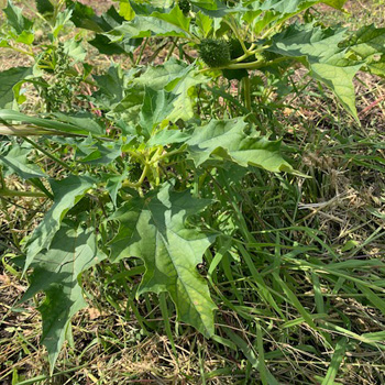
Introduction: Sudden onset of reduced consciousness, psychomotor agitation and mydriasis are all indicative of an anticholinergic toxidrome. It is important to note that numerous drugs, as well as certain herbs and plants, possess anticholinergic properties.
Case description: An 84-year-old female patient had sudden nocturnal onset of uncoordinated hand movements and altered mental status. Shortly after, the patient’s 83-year-old husband developed symptoms of dysarthria, gait ataxia, vertigo, and delirium.
Conclusion: Anticholinergic syndrome consists of a combination of central and peripheral anticholinergic symptoms. Physostigmine given intravenously resulted in rapid reversal of symptoms. Thorn apple seeds had been accidentally ingested and were identified as the cause.
|
Views: 63
HTML: 8
PDF: 42
|
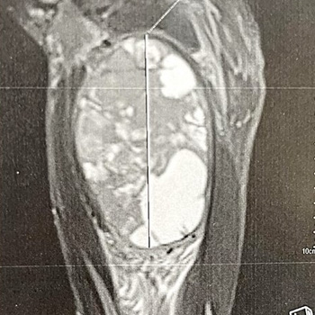
Background: Patients with neurofibromatosis type I (NF1) have an increased risk of developing soft-tissue sarcomas, particularly those related to the nervous system. Epithelioid sarcoma (ES) is an exceptionally rare subtype of soft-tissue sarcoma, with limited knowledge about its clinical presentation and optimal management in NF1. This report aims to provide insights into the characteristics and outcomes of ES in NF1 patients.
Case description: A 37-year-old man with a history of NF1 presented with a progressively worsening mass on his right inner thigh. An MRI scan revealed a well-defined tissue mass originating from the adductor magnus muscle, later confirmed as ES through histopathology and immunohistochemistry. Considering poor local and general prognosis, the multidisciplinary team recommended salvage hip disarticulation, however the patient refused and opted for palliative marginal resection to reduce the tumour size. The patient’s condition declined rapidly, and he succumbed six days after the surgery.
Conclusion: This case highlights the rarity of ES in NF1 patients and underscores the potential for malignant tumour development in this population. Further research is needed to improve our understanding and management of sarcomas in the context of NF1.
|
Views: 60
HTML: 11
PDF: 48
|
We present a 30-year-old male who sustained a mild traumatic brain injury and then was intubated due to deterioration of consciousness. A head CT scan revealed mild brain oedema, a fractured nasal bone and mild left thoracic wall haematoma. Despite complete clinical and radiological normalisation within 36 hours, he failed to wean off the ventilator. The patient was found to have subtle bulbar manifestations including dysphonia, dysarthria, and dysphagia, with recurrent left lung collapse. He responded to an empirical pyridostigmine trial despite negative biochemical tests for myasthenia gravis (MG). The patient was weaned successfully from the ventilator, transferred to a long-term care facility, and then discharged home. Classic symptoms and signs of a disease may be absent, but the presence of dysarthria, dysphagia, transient vocal cord palsy, nasal speech, absent gag reflex and respiratory failure in difficult-to-wean patients, with no definitive diagnosis, may warrant an empirical trial of therapy for suspected MG and for the benefit of any doubt.
|
Views: 44
HTML: 12
PDF: 42
|
Introduction: Rectus sheath haematoma (RSH) has become increasingly common but is often underdiagnosed. Prompt diagnosis will avoid unnecessary investigations and procedures, resulting in early treatment and a better outcome.
Case description: We described a case of a spontaneous RSH with intraperitoneal extension and formation of a vesico-haematoma fistula, which was initially misdiagnosed as a urinary tract infection. The diagnosis was made ten days after admission, when a CT scan showed an over-16 cm RSH with intraperitoneal extension, bladder perforation and a vesico-haematoma fistula. The patient was managed conservatively.
Discussion: RSH accounts for less than 2% of acute abdomen cases and is often unrecognised. Its presentation can mimic other intra-abdominal pathologies, and the diagnosis is often delayed or missed. Complications can arise from an RSH although it is generally viewed as a self-limiting condition.
Conclusion: RSH has become increasingly common, and we would like to highlight the need to include abdominal wall pathologies in the initial differential diagnoses of acute abdomen to avoid delay in diagnosis.
|
Views: 51
HTML: 6
PDF: 37
|
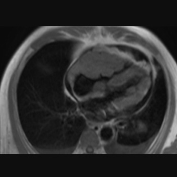
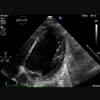
Background: This study presents a patient diagnosed with tricuspid valvular stenosis due to right ventricular lymphoma, who was treated successfully.
Case presentation: A 66-year-old man with a history of worsening shortness of breath during activity for the last three weeks sought medical attention. The patient later experienced swelling in the extremities, fluid build-up around the lungs and abdominal fluid accumulation, with no reported chest pain, fever, or weight loss. An echocardiogram found a mass in the lateral wall near the tricuspid valve of the right ventricle, leading to moderate tricuspid stenosis. The cardiac magnetic resonance imaging (MRI) revealed a lumpy, poorly defined mass that invaded the heart muscle and displayed varied enhancement after contrast administration. Suspicion arose for a malignant tumour or metastatic lesion due to its features and contrast uptake capability. A percutaneous biopsy was carried out on the mass in the right ventricle to confirm the diagnosis. The pathology report indicated a diagnosis of non-Hodgkin’s lymphoma. After being diagnosed, the patient underwent chemotherapy using the R-CHOP regimen. Over time the symptoms improved, and echocardiograms revealed a decrease in the size of the tumour. After undergoing six rounds of chemotherapy, a cardiac MRI four months later showed no signs of a tumour. After that, the patient resumed their regular activities.
Conclusion: Right ventricular tumours are mostly malignant lesions and often have an inferior prognosis. Early diagnosis with imaging techniques and myocardial biopsy is necessary to deliver life-saving treatment quickly.
|
Views: 43
HTML: 8
PDF: 32
|
Pleuroperitoneal leak as a cause of pleural effusions in peritoneal dialysis is a rare but important complication to consider in continuous ambulatory peritoneal dialysis (CAPD) patients presenting with recurrent progressive dyspnoea. Generally, these effusions are unilateral and right-sided, resulting in shortness of breath and reduced ultrafiltration volume, which are initially managed by peritoneal rest. We describe a case of bilateral pleural effusions in a 57-year-old female on chronic CAPD who developed recurrent progressive dyspnoea but maintained adequate dialysis output. A chest radiograph revealed bilateral pleural effusions with high glucose content, and scintigraphy confirmed the existence of a definite pleuroperitoneal communication. She was managed by temporary substitution to haemodialysis, followed by suturing of the shunt and successful video-assisted thoracoscopic surgery (VATS) pleurodesis with an aldehyde-based surgical glue. Unexplained recurring dyspnoea in chronic CAPD should raise the suspicion of a possible pleuroperitoneal leak, even in patients without an apparent loss of ultrafiltration. Pleurodesis using an aldehyde-based adhesive was effective and tolerated well by our patient and may be considered in managing cases of recurrent pleural effusion.
|
Views: 82
HTML: 16
PDF: 72
|
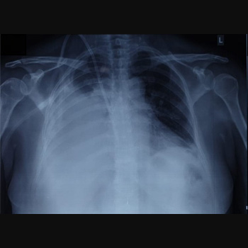
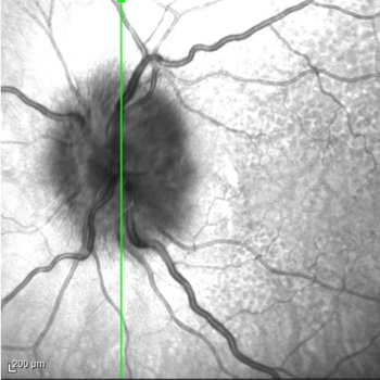
Background: eosinophilic granulomatosis with polyangiitis (EGPA) is a rare multisystem inflammatory disease characterized by asthma, eosinophilia and granulomatous or vasculitic involvement of various organs. While the eye is uncommonly affected in patients with EGPA, multiple ophthalmic manifestations have been reported, which can result in serious visual impairment without timely treatment.
Case report: we report the case of a 79-year-old woman with a history of asthma and nasal polyps who presented with low-grade fever, mild alteration of mental status, and fatigue. Chest X-ray revealed bilateral interstitial infiltrates. Lab tests showed elevated C-reactive protein level and eosinophilia (eosinophil count, 4.6 x10⁹ cells/l); blood cultures and parasitological examination of stools tested negative. Four days after presentation, the patient reported sudden and severe blurring of vision in her left eye. Ophthalmological examination revealed bilateral swollen optic disc and visual field loss, more severe in the left eye. A diagnosis of EGPA complicated by arteritic anterior ischaemic optic neuropathy (A-AION) was proposed, while an alternative or concurrent diagnosis of giant cell arteritis was ruled out based on clinical picture.
Immunosuppressive treatment with high-dose intravenous glucocorticoids was promptly started. The patient’s visual defect did not improve; however, two months later, no worsening was registered on ophthalmic reassessment.
Conclusions: A-AION is an infrequent but severe manifestation of EGPA, requiring prompt diagnosis and emergency-level glucocorticoid therapy to prevent any further vision loss. Disease awareness and a multidisciplinary approach are crucial to expedite diagnostic work-up and effective management of EGPA-related ocular complications.
|
Views: 117
HTML: 28
PDF: 70
|
Introduction: Combination-based adjuvant chemotherapy utilising capecitabine and oxaliplatin is widely used in gastric cancer treatment. Rare but severe cardiac events such as prolonged QT, cardiac arrest and cardiogenic shock can result from their use.
Case description: A 45-year-old female with gastric adenocarcinoma was started on capecitabine-oxaliplatin chemotherapy one week before presenting to the emergency department with weakness. Blood pressure was 78/56 mmHg, heart rate 140 bpm and oxygen saturation 85%. She became unresponsive with pulseless ventricular fibrillation; CPR was initiated with immediate intubation. She received two shocks with a return of spontaneous circulation. Laboratory tests revealed serum potassium (3.1 mmol/l), magnesium (1.1 mg/dl) and troponin (0.46 ng/ml). An EKG revealed sinus tachycardia with a prolonged QT interval (556 ms). The combined effects of capecitabine, oxaliplatin and electrolyte abnormalities likely contributed to the QT prolongation. An echocardiogram demonstrated an ejection fraction of 10%–15%. An emergent right-heart catheterisation showed right atrial pressure of 10 mmHg and pulmonary artery pressure of 30/18 mmHg; cardiac output and index were not recorded. An intra-aortic balloon pump was placed, and she was admitted to the ICU for cardiogenic shock requiring norepinephrine, vasopressin and dobutamine. A repeat echocardiogram showed a significantly improved ejection fraction of 65%, and she was discharged.
Discussion: Capecitabine and oxaliplatin cardiotoxicity is an exceedingly rare occurrence, with both drugs reported to cause QT prolongation.
Conclusion: Healthcare providers must recognise the QT prolongation effects of capecitabine and oxaliplatin, leading to life-threatening cardiac arrhythmias.
|
Views: 196
HTML: 50
PDF: 212
|
Background: Tirzepatide is a novel glucagon-like peptide 1/glucose-dependent insulinotropic peptide (GLP-1/GIP) receptor agonist. It was recently approved for diabetes control and weight reduction in non-diabetic patients.
Case description: We report the first case of ketoacidosis after the use of tirzepatide in an obese non-diabetic patient, secondary to the possibility of starvation ketoacidosis and insulin resistance.
Conclusion: The dual-acting GLP-1 and GIP receptor agonists, tirzepatide, can induce ketoacidosis in obese non-diabetic patients.
|
Views: 68
HTML: 8
PDF: 67
|
Introduction: Most pregnancies in women after a kidney transplant result in a live birth, but kidney functions should be stable for one year before conception. For immunosuppression modification occurring before pregnancy, azathioprine is used because it is considered safe for major congenital malformations during pregnancy. However, there may be an association between exposure to azathioprine during pregnancy and the onset of an unusual, early and severe form of intrahepatic cholestasis.
Case description: A young patient with a twin pregnancy after a second kidney transplant experienced intrahepatic cholestasis. There was a wide range of differential diagnosis. A battery of tests was requested including autoimmune markers, virology, and imaging. The conclusion that azathioprine was contributing to intrahepatic cholestasis with pregnancy was reached after exclusion of all other differentials.
Conclusions: Complications of pregnancy after a kidney transplant include hypertension, pre-eclampsia, deterioration of graft function up to rejection, but also unusual side effects of immunosuppression medication.

|
Views: 59
HTML: 15
PDF: 56
|
Kocuria kristinae is a Gram-positive commensal bacterium, rarely responsible for infection in immunocompromised patients.
A 29-year-old woman affected by intestinal pseudo-obstruction and requiring home parenteral nutrition, was hospitalised for fever and shivering during the infusion through a long-term central venous catheter (CVC).
Blood cultures were positive for K. kristinae infection. At a chest CT scan, two partially cavitated nodular lesions were evidenced. Meropenem antibiotic therapy was used locally and systemically, resulting in catheter use restoration.
A chest CT scan two months later at follow-up showed two centimetric, fibrotic and disventilatory areas replacing the previous nodular thickenings.
Kokuria kristinae was responsible for haematogenous pulmonary involvement with excavated nodules, requiring a differential diagnosis. Moreover, in the case of a CVC infection, in addition to the risk of right endocarditis, haematogenous pneumonia must also be considered.
|
Views: 99
HTML: 12
PDF: 95
|
Introduction: A case of ocular bartonellosis under anti-tumour necrosis factor treatment is described.
Case description: A 29-year-old woman with psoriasis who had been on certolizumab treatment was examined with a left visual deterioration following a fever bout, malaise, and placoid erythematous rashes on her neck. As there was acute anterior uveitis in her left eye, it was recommended to stop certolizumab treatment for a possible infectious aetiology. However, her physician elected to continue the certolizumab treatment. Ten days later, the patient noticed further visual decline despite the topical steroid treatment. This time, there were scattered yellow-white small retinitis foci at the left posterior pole. Infectious agents were searched and while Bartonella henselae antibodies were negative for immunoglobulin M, the immunoglobulin G titre was 1/80. Clinical findings were improved with the systemic treatment of oral trimethoprim-sulfamethoxazole (160/800 mg twice daily for six weeks) and azithromycin (500 mg once daily for two weeks).
Discussion: Though extremely rare, ocular bartonellosis should be kept in mind in patients on anti-tumour necrosis factor treatment as rapid and accurate diagnosis may end up with an excellent visual outcome and full recovery.
|
Views: 80
HTML: 9
PDF: 70
|
Introduction: Pott’s puffy tumour is a rare entity defined by the presence of a subperiosteal abscess of the frontal bone associated with frontal osteomyelitis. Several predisposing conditions can lead to this entity, such as frontal sinusitis.
Case description: We report the case of a 15-year-old patient who presented to the emergency department for headache, fever and forehead swelling. Computed tomography revealed severe pansinusitis complicated by a subperiosteal abscess associated with frontal osteomyelitis, leading to the diagnosis of Pott’s puffy tumour. The management combined intravenous antibiotics and surgical drainage of both the sinusitis and subperiosteal abscess.
Discussion: Pott’s puffy tumour represents a rare but serious complication of frontal sinusitis. Clinicians should be aware of this potential complication as the diagnosis can be challenging at an early stage but may influence the subsequent prognosis.

|
Views: 107
HTML: 17
PDF: 59
|
Peritonitis, the inflammation of the protective membrane surrounding parts of the abdominal organs, is a common clinical pathology with multifactorial aetiologies. While bacterial infections are well-recognised as a cause of peritonitis, fungal infections remain relatively uncommon especially Saccharomyces cerevisiae, which is commonly used for breadmaking and as a nutritional supplement. This fungus has been reported to induce peritonitis in patients on peritoneal dialysis. However, it has never been reported as secondary to percutaneous endoscopic gastrostomy (PEG) tube insertion in immunocompromised patients. We present a 64-year-old female with a history of human immunodeficiency virus (HIV) who developed S. cerevisiae peritonitis following PEG tube insertion. The case highlights the importance of considering rare organisms when treating immunocompromised patients with peritonitis, especially after gastrointestinal tract penetration or peritoneal membrane disruption.

|
Views: 157
HTML: 39
PDF: 88
|
Chronic lymphocytic leukaemia (CLL) is a lymphoproliferative disorder characterised by an accumulation of monoclonal B lymphocytes, with an increased risk of secondary cancers. The coexistence of CLL and chronic myeloid leukaemia (CML) is a rare phenomenon, with three main types being classified: CML preceding CLL, CLL preceding CML and simultaneous occurrence. The coexistence of these chronic leukaemias poses a complex clinical challenge, with the underlying mechanisms of their association remaining enigmatic. Here, we present a report of an elderly male with a long history of CLL, who was subsequently diagnosed with secondary CML.
|
Views: 164
HTML: 23
PDF: 96
|
Background: This report presents the influence of immunosuppression by new rheumatological therapies on hepatitis E virus infection in a 54-year-old male patient with an anti-synthetase syndrome and treatment with methotrexate and rituximab.
Case description: The patient arrived at the Emergency Department with epigastric pain, vomiting and dark urine. Initial examination revealed signs of inflammation and hepatic dysfunction. Subsequent laboratory tests and imaging confirmed acute hepatitis E infection in the context of recent initiation of rituximab therapy. Despite initial suspicion of pancreatitis, subsequent investigations ruled out pancreatic involvement. Treatment with ribavirin, along with supportive measures, led to significant clinical improvement with resolution of jaundice, ascites, and oedema.
Conclusions: This case underscores the importance of considering hepatitis E in patients with autoimmune conditions, especially when initiating immunosuppressive therapies, a situation that is not well described in scientific literature and is increasingly common, necessitating proper recognition.
|
Views: 144
HTML: 18
PDF: 104
|
Ulcerative colitis (UC) is an autoimmune disease associated with both intestinal and extraintestinal manifestations. The latter may include heart complications, such as myopericarditis leading to life-threatening arrythmias. Nowadays, UC is commonly treated with biologic medications and infliximab is the first line therapy in an outpatient setting, while it is also used as rescue therapy in acute severe UC. However, it has been associated with severe immunosuppression, cytomegalovirus (CMV) reactivation and drug-induced hepatitis. We report a case of UC flare in a biologic naïve patient admitted with myopericarditis, which was further complicated by positive CMV biopsies and infliximab-induced transaminitis.
| 2.1 = | 1.730 Cit. to date |
| 842 Docs. to date |


Publisher
Official Journal of the
European Federation of Internal Medicine
www.efim.org
Publisher: SMC media Srl
Via Giovenale, 7 - 20136 Milan - Italy
P.IVA 07626490960
info@ejcrim.com
www.ejcrim.com - ISSN: 2284-2594 - © EFIM 2014-2023, Published by SMC Media srl, Italy - Privacy policy

