DOI: 10.12890/2024_004760
Heterotopic splenic tissue mimicking metastases on magnetic resonance imaging
Authors
 cli4d@live.com
Keywords
Splenosis, heterotopic spleen, prostate, cancer, adenocarcinomaAbstract
Background: Heterotopic splenic tissue can occur following splenectomy and is typically asymptomatic, often discovered incidentally during imaging for other conditions. This benign condition may mimic malignant processes, posing diagnostic challenges especially in patients with a history of cancer or concurrent malignancy.
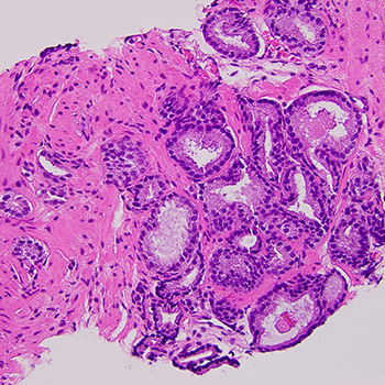
Case description: We report the case of a 60-year-old male with a history of well-controlled hypertension and a splenectomy following a traumatic injury at age 7. The patient underwent routine screening which revealed elevated prostate-specific antigen (PSA) levels. Subsequent magnetic resonance imaging (MRI) identified suspicious lesions in the prostate and a left lower quadrant mass. Prostate biopsy confirmed an adenocarcinoma with a Gleason score of 6, while biopsy of the abdominal mass revealed heterotopic splenic tissue. The management strategy included active surveillance for prostate cancer, considering the tumour’s low aggressiveness and the benign nature of the splenic tissue.
Conclusions: This case highlights the importance of considering heterotopic splenic tissue in differential diagnosis when evaluating abdominal masses in patients with a history of splenectomy. Accurate diagnosis through careful imaging and biopsy is crucial to avoid misdiagnosis and unnecessary treatments, particularly in patients with concurrent malignancies.
References
- Fremont RD, Rice TW. Splenosis: a review. South Med J 2007;100:589–593.
- Kawachi MH, Bahnson RR, Barry M, Busby JE, Carroll PR, Carter HB, et al. NCCN clinical practice guidelines in oncology: prostate cancer early detection. J Natl Compr Canc Netw 2010;8:240–262.
- Tandon YK, Coppa CP, Purysko AS. Splenosis: a great mimicker of neoplastic disease. Abdom Radiol (NY) 2018;43:3054–3059.
- Vernuccio F, Dimarco M, Porrello G, Cannella R, Cusmà S, Midiri M, et al. Abdominal splenosis and its differential diagnoses: what the radiologist needs to know. Curr Probl Diagn Radiol 2021;50:229–235.
- Lin WC, Lee RC, Chiang JH, Wei CJ, Chu LS, Liu RS, et al. MR features of abdominal splenosis. AJR Am J Roentgenol 2003;180:493–496.
- Lake ST, Johnson PT, Kawamoto S, Hruban RH, Fishman EK. CT of splenosis: patterns and pitfalls. AJR Am J Roentgenol 2012;199:W686–693.
- Mariani G, Bruselli L, Kuwert T, Kim EE, Flotats A, Israel O, et al. A review on the clinical uses of SPECT/CT. Eur J Nucl Med Mol Imaging 2010;37:1959-1985.
- March B, Koufogiannis G, Louie-Johnsun M. Management and outcomes of Gleason six prostate cancer detected on needle biopsy: a single-surgeon experience over 6 years. Prostate Int 2017;5:139–142.
- Moore CM, King LE, Withington J, Amin MB, Andrews M, Briers E, et al. (2023). Best current practice and research priorities in active surveillance for prostate cancer – a report of a Movember International Consensus meeting. Eur Urol Oncol 2023;6:160–182.