DOI: 10.12890/2024_004710
Can lesions in the right basal ganglia cause aphasia? Crossed aphasia in a right-handed patient
Authors
 mjstrindade@gmail.com
Keywords
Crossed aphasia, crossed aphasia in dextral, aphasiaAbstract
Background: Aphasia is a common neurocognitive disorder caused by impaired speech and language, with stroke being the most frequent cause. The neuroanatomical mechanism underlying this condition is not yet fully understood.
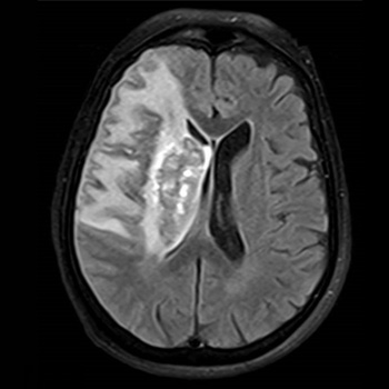
Case description: This case describes a 74-year-old Caucasian woman admitted with a clinical picture of right total anterior circulation infarct (TACI) and aphasia, scoring 17 on the National Institutes of Health Stroke Scale. Neuroimaging showed a large cortico-subcortical frontotemporoparietal and insular infarct involving the basal ganglia of the right hemisphere and bilateral focal atherosclerotic stenosis on the M1 segment of the middle cerebral artery. There was no left hemispheric lesion or abnormal electric activity on the electroencephalogram. A formal evaluation was compatible with transcortical motor aphasia. The aetiological study revealed atrial fibrillation, and the case was admitted as an ischaemic stroke of undetermined aetiology with two possible causes – intracranial atherosclerotic stenosis or atrial fibrillation.
Conclusion: Our patient fulfilled all the formal criteria for crossed aphasia in dextral (CAD): aphasia, a lesion in the right hemisphere coupled with the structural integrity of the left hemisphere, an established preference for right-hand use without a familial history of left-handedness individuals, and an absence of brain damage in childhood. Our patient’s case adds to the evidence that deep structures – alone or in combination with cortical structures – are primarily affected in CAD.
References
- Ngo D, Aftab G, Madhavan A, Bukhari A. Idiopathic pulmonary vein thrombosis treated with apixaban. Respirol Case Rep 2021;9:e00803.
- Selvidge SD, Gavant ML. Idiopathic pulmonary vein thrombosis: detection by CT and MR imaging. AJR Am J Roentgenol 1999;172:1639–1641.
- Komatsu S, Kamata T, Imai A, Miyaji K, Ohara T, Takewa M, et al. Idiopathic pulmonary vein thrombosis complicated with old myocardial infarction detected by multidetector row computed tomography. J Cardiol Cases 2011;3:e94–e97.
- Goddard SA, Tran DQ, Chan MF, Honda MN, Weidenhaft MC, Triche BL. Pulmonary vein thrombosis in COVID-19. Chest 2021;159:e361–e364.
- Mumoli N, Cei M. Idiopathic pulmonary vein thrombosis. J Emerg Med 2012;42:182–183.