EJCRIM 2023 CiteScore
| 2.1 = | 1.762 Cit. to date |
| 842 Docs. to date |
Last updated on 05 May, 2024
Updated monthly
Updated monthly
Powered by

|
Views: 662
HTML: 140
PDF: 305
|
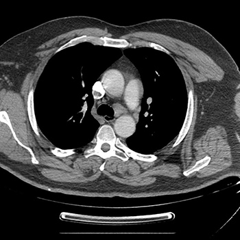
Mediastinal tumours can be incidental findings on chest x-ray or present with systemic symptoms and/or direct effect of the mediastinal mass. We report the case of a woman with symptomatic thymoma B1 and simultaneous thymus tuberculosis.
|
Views: 470
HTML: 82
PDF: 332
|
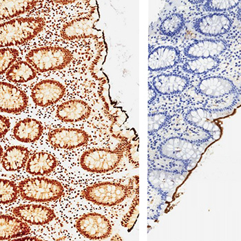
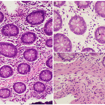
Serrated polyposis syndrome (SPS) is the most common form of polyposis syndrome and has been shown to increase the risk of colorectal cancer (CRC). The genetic pathway of CRC in SPS is different from the classic adenomatous polyposis coli (APC) pathway, which accounts for 70–80% of cases of CRC. Most commonly, SPS mutations include BRAF and KRAS, with activation of the RAS-RAF-MAP kinase pathway involved in the pathogenesis of serrated lesions. We present a rare case of SPS in a 32-year-old woman with MSH6 and SMARCA4 variants, which have not previously been reported in the literature.
|
Views: 578
HTML: 63
PDF: 344
|
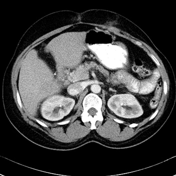
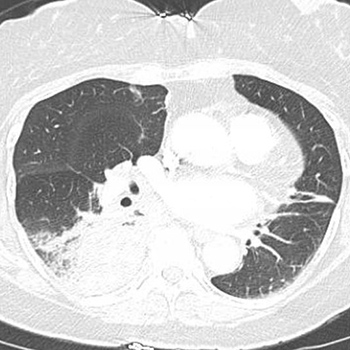
SARS-CoV-2 causes severe acute respiratory distress and other clinical complications such as thromboembolic events and gastrointestinal tract disorders, which generally present with abdominal pain. In the case report, we describe a patient who had severe viral necrotizing pancreatitis associated with COVID-19 infection.
|
Views: 536
HTML: 106
PDF: 253
|
Giant cell arteritis is a medical emergency as severe, irreversible complications may occur if it is not treated in a timely manner. However, in daily practice early diagnosis can be challenging. We report the case of a 70-year-old woman who presented with multiple ischaemic cerebral vascular accidents related to newly diagnosed giant cell arteritis. Review of her charts revealed a substantial delay from the onset of symptoms to diagnosis. This case demonstrates the need for additional efforts to reduce delay in referring patients with giant cell arteritis and the need to implement fast-track clinics to prevent serious complications.
|
Views: 639
HTML: 368
PDF: 438
|
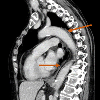
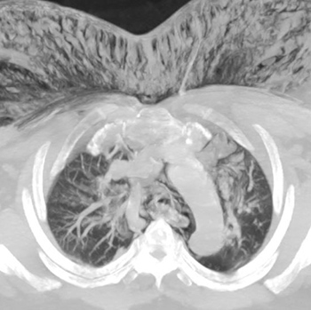
Tracheal tears are an uncommon phenomenon that can occur due to infection, blunt or penetrating trauma or iatrogenic causes secondary to endotracheal intubation or procedures such as bronchoscopy. Post-intubation tracheal laceration is a very rare yet serious complication with high morbidity and mortality rates. Here, we report the case of a 53-year-old woman with a medical history of hypertension who presented with complaints of facial swelling after undergoing arthroscopic debridement of the coracoacromial ligament as well as partial resection of the acromion for impingement of the right shoulder under general anaesthesia. The patient was found to have extensive pneumomediastinum, subcutaneous emphysema, and a large tracheal tear. We aim to highlight this rare complication of endotracheal intubation, discuss the presenting signs and symptoms, and explore the various management options.
|
Views: 638
HTML: 180
PDF: 281
|
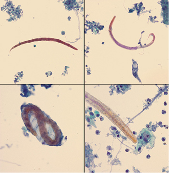
Ciliocytophthoria is a phenomenon where degenerated cells in infections or malignancy can present as ciliated cells on microscopy and so may be confused with ciliated parasitic infection. We present an interesting case of recurrent shortness of breath, misdiagnosed as chronic obstructive pulmonary disease exacerbations leading to unnecessary exposure to antimicrobials and steroids. The case was diagnosed as Strongyloides hyper-infection syndrome. Another finding worth mentioning was that ciliated cells noted on broncho-alveolar lavage were thought to be a co-infection with Balantidium coli but were later confirmed as ciliocytophthoria.
|
Views: 913
HTML: 155
PDF: 477
|
Kluver-Bucy syndrome (KBS) is a characterized by a group of cognitive dysfunctions that include hypersexuality, placidity, hyperorality, memory deficits and hypermetamorphosis. This syndrome is often seen in pathological states that destroy the temporal lobes, normally bilaterally. Herpes simplex encephalitis (HSE) is one of the causes of KBS, as the herpes virus can cause dysfunction/destruction of the temporal lobes. KBS is a very rare syndrome, with just a few cases described in the literature. We present the case of a 21-year-old-man who was diagnosis with KBS after HSE.
|
Views: 876
HTML: 64
PDF: 337
|
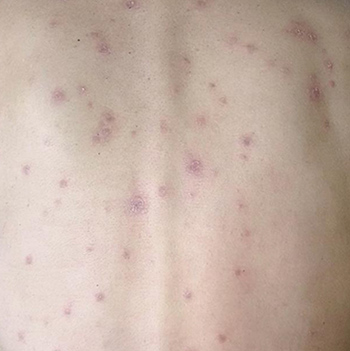
Erythema nodosum (EN) is an inflammatory condition of the subcutaneous fat and has been reported in patients with haematological malignancies (lymphomas) or solid tumours. Lung cancer is the most common cause of paraneoplastic syndrome. We report a case of EN occurring as a paraneoplastic disease.
A 48-year-old Tunisian woman, a non-smoker with no relevant medical history, presented with painful, erythematous, firm nodules on her legs with ankle swelling. The patient did not report any other symptoms. There were no abnormalities on examination except for moderate fever. An extensive infectious and immunological investigation was negative. Antistreptolysin antibodies were undetectable. Chest radiography showed a focal opacity in the right lung and a CT scan revealed a mass in the lower right pulmonary lobe with hilar and mediastinal lymphadenopathies, a nodule in the right adrenal gland, condensation in the iliac bone and multiple bilateral nodular cerebral expansive processes. Bronchial biopsies revealed a primitive and moderately differentiated adenocarcinoma. No argument for tuberculosis or sarcoidosis was found.
|
Views: 514
HTML: 109
PDF: 249
|
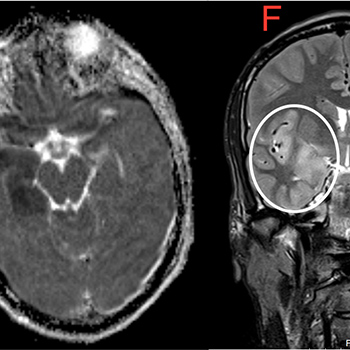
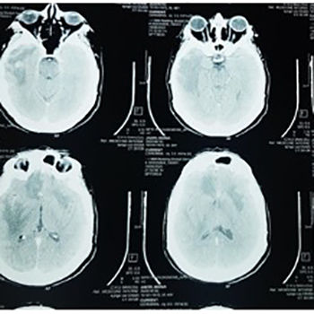
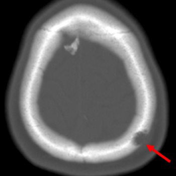
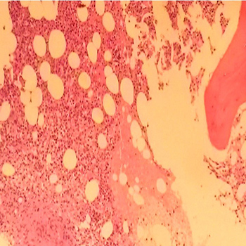
Eosinophilic granuloma is a localized, non-invasive form of Langerhans cell histiocytosis. It usually develops in the long bones and is more frequent in children under the age of 10 years. It is very rare in adults.
We present the case of a young woman admitted to hospital for persistent refractory left parietal headache, later revealed to be caused by an eosinophilic granuloma.
|
Views: 721
HTML: 992
PDF: 205
|
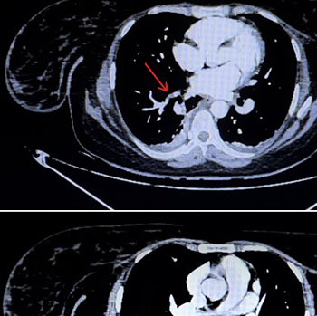
In secondary syphilis, Treponema pallidum can spread to the lungs. A new case is described of a patient with multiple excavated subpleural pulmonary nodules, a rare localization of secondary syphilis. Despite the numerous pulmonary samples analysed in the literature, T. pallidum is rarely visualised directly on bronchial fibroscopy or biopsy. The diagnosis of secondary syphilis is suspected from historical and physical findings and confirmed by high values obtained from non-treponemal tests.
|
Views: 784
HTML: 62
PDF: 427
|
The prevalence of venous thromboembolism (VTE) in COVID-19 patients is higher than in non-COVID-19 patients. Since the beginning of the pandemic, deep vein thrombosis, myocardial infarction, ischaemic stroke and pulmonary embolism (PE) have been reported in patients with COVID-19. D-dimer levels are now routinely measured in hospitalized patients so that prophylaxis can be initiated. However, a standardized protocol for prophylaxis has yet to be developed for pregnant women with COVID-19, who have an increased risk of VTE. We describe the case of a young primigravida woman with a positive COVID RT-PCR test who developed PE despite receiving adequate prophylaxis.
|
Views: 877
HTML: 306
PDF: 497
|
Coronavirus disease 2019 (COVID-19) is caused by severe acute respiratory syndrome coronavirus 2 (SARS-CoV-2). Clinical manifestations are diverse and can vary from mild respiratory symptoms to severe hypoxic respiratory failure. In severe cases, infection can cause gastrointestinal, renal, cardiac, neurological and haematological complications and result in multi-organ failure. There are very few reports of parapneumonic effusion in patients with COVID-19. We describe two patients with COVID-19 who had loculated empyema and discuss the clinical course and therapeutic options.
|
Views: 613
HTML: 62
PDF: 363
|
The authors present a case of purulent pericarditis probably secondary to respiratory infection, a rare entity in the antibiotic era. Pericardial fluid analysis identified streptococci and oral anaerobes as the causative agents. A prolonged and complicated diagnostic and therapeutic course, which included a long stay in the intensive care unit, is described, and a review of purulent pericarditis provided. Pericardial effusion, particularly in the setting of concomitant respiratory infection and immunocompromise or other risk factors, should raise the suspicion of bacterial pericarditis and prompt its timely diagnosis and treatment. Purulent pericarditis can be lethal and has potentially severe complications, so adequate antimicrobial therapy and source control are key.
|
Views: 883
HTML: 60
PDF: 480
|
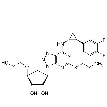
Ticagrelor is a directly acting cyclopentyltriazolo-pyrimidine which does not require conversion into an active metabolite. It inhibits the P2Y12 receptors on platelets reversibly. Unlike clopidogrel and prasugrel, resistance to ticagrelor is rarely reported. Various mechanisms have been proposed for this resistance. The case of a 62-year-old man with diabetes who had undergone index percutaneous coronary intervention (PCI) 22 days previously is described. The patient presented to us with stent thrombosis. His primary PCI was successfully carried out with a drug-eluting stent. He showed resistance to ticagrelor on thromboelastography platelet mapping. He responded well to prasugrel (another P2Y12 inhibitor) in combination with aspirin.
|
Views: 769
HTML: 651
PDF: 301
|
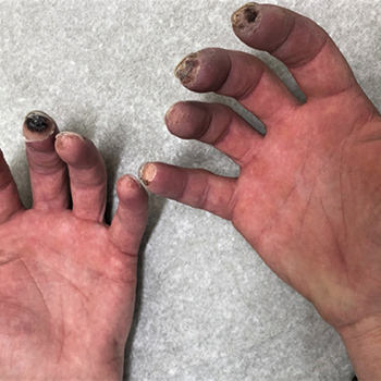
Paraneoplastic Raynaud’s phenomenon has often been reported in leukaemia, lymphoma and sarcoma. Nevertheless, an association with lung cancer is not frequently reported in the literature even though lung cancer is a common malignancy. We present a case of paraneoplastic Raynaud’s phenomenon as the presenting feature of underlying lung malignancy.
|
Views: 578
HTML: 68
PDF: 330
|
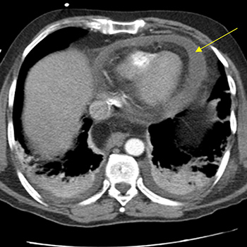
Sarcoidosis is a systemic granulomatous disease in which medullary involvement is a rare extrapulmonary manifestation. We present the case of a 37-year-old man with right abdominal and dorso-lumbar pain lasting for months. Computerized tomography showed renal microlithiasis and retroperitoneal, hilar and mediastinal adenopathies. Laboratory results showed an elevated erythrocyte sedimentation rate, IgG, ?2-microgobulin and angiotensin-conversion enzyme, serum calcium in the upper limit and hypercalciuria. There was a slight elevation of the CD4/CD8 ratio in bronchoalveolar lavage, without lymphocytic alveolitis. An endobronchial ganglion biopsy was inconclusive. A positron emission tomography scan demonstrated supra and infra-diaphragmatic, splenic and medullary involvement, suggesting lymphoproliferative disease (LPD). A bone marrow biopsy (BMB) revealed sarcoid-like epithelioid cell granulomas, excluding LPD. Sarcoidosis was assumed and corticosteroids were started. Although cytopenias were not present, the extensive ganglion, splenic and medullary involvement made LPD exclusion imperative, while BMB allowed for a definitive diagnosis.
|
Views: 537
HTML: 72
PDF: 308
|
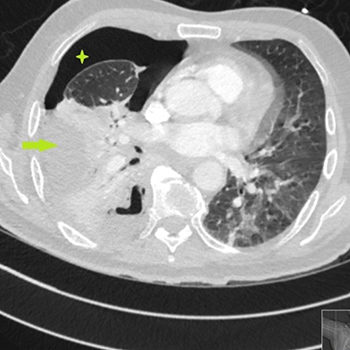
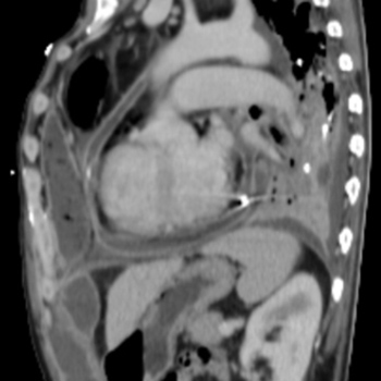
Cardiac tamponade is a life-threatening medical emergency and can arise in many clinical situations.
We present the case of a 59-year-old man with adrenoleukodystrophy and Addison’s disease who was admitted to the emergency department with severe abdominal pain that turned out to be cardiac tamponade of unknown aetiology.
An association between cardiac tamponade and Addison’s disease has been reported in the literature, so this aetiology should be considered in the differential diagnosis for patients presenting with unexplained cardiac tamponade.
|
Views: 490
HTML: 110
PDF: 223
|
We present the case of an HIV-positive patient admitted because of costal pain secondary to neoplasia. During investigations, a colonoscopy showed non-specific ulcerations. Histological examination resulted in a diagnosis of intestinal spirochetosis. This infection can be asymptomatic or cause non-specific symptoms such as diarrhoea or abdominal pain. Intestinal spirochetosis should be included in the differential diagnosis of colon lesions in patients with HIV infection.
|
Views: 764
HTML: 904
PDF: 415
|
Reactive thrombocytosis after splenectomy is a feared cause of thrombosis throughout the arterial and venous system. There are many causes of splenomegaly, ranging from cirrhosis to lymphoma to hereditary spherocytosis. In this report, we will discuss a case of reactive thrombocytosis after splenectomy in a patient with hereditary spherocytosis. Splenomegaly is a relatively common finding in HD patients, causing extravascular haemolysis and thus leading to haemolytic anaemia. Splenectomy is usually considered when patients start to manifest severe symptoms such as abdominal pain, jaundice or worsening liver function tests. Our patient was a good surgical candidate and successfully underwent splenectomy but afterwards developed arterial and venous thrombosis due to reactive thrombocytosis. An extensive hypercoagulable work-up was unremarkable. The patient was started on hydroxyurea and anticoagulation with eventual improvement of platelet levels.
|
Views: 562
HTML: 108
PDF: 309
|
Introduction: Aortitis is seen in a wide variety of diseases. It was rarely found in the past but this is changing because of new imaging techniques.
Case description: We present the case of a 45-year-old man who was found on thyroid ultrasound to have infrarenal aortitis and pathological lymphadenopathies in different locations. After an exhaustive diagnostic process, tuberculous aortitis, an infrequent manifestation of extrapulmonary tuberculosis, was diagnosed. The condition resolved after a 6-month course of antibiotics and a 6-week course of corticosteroids.
Conclusion: Tuberculous aortitis is an atypical manifestation of Mycobacterium tuberculosis infection. The absence of typical symptoms and the difficulty of isolating the microorganism makes its diagnosis difficult. Therefore, clinical suspicion, microbiological tests and imaging are key for reaching the diagnosis and starting treatment for a serious disease that can cause aortic aneurysm and dissection.
|
Views: 588
HTML: 76
PDF: 256
|
Eosinophilic colitis is a rare heterogeneous inflammatory disorder. The pathogenesis is not well understood even though it seems to be multifactorial, with hypersensitivity as a major contributor. The clinical presentation depends on the eosinophilic infiltration of different sections within the gastrointestinal tract. Diagnosis is based on the presence of peripheral eosinophilia and histopathological evidence of colon wall eosinophilic infiltration.
The authors present the case of a woman with a predominantly subserous pattern of eosinophilic colitis potentially triggered by intake of an Ulmus rubra-rich product.
|
Views: 556
HTML: 91
PDF: 293
|
Pulmonary coccidioidomycosis and pulmonary actinomycosis are unheard of as co-pathogens. Infection with these organisms on their own can mimic lung cancer, thus presenting a diagnostic challenge. We present the case of a 75-year-old woman presenting with haemoptysis with a chest CT chest finding of a lung mass suggestive of lung cancer. A diagnosis of concomitant infection by Coccidioides posadasii/immitis and Actinomyces odontolyticus was made based on culture and histopathology results. The patient was successfully treated with a combination of antifungal and antibacterial therapy. This is the first reported case of co-infection by these two microorganisms.
| 2.1 = | 1.762 Cit. to date |
| 842 Docs. to date |


Publisher
Official Journal of the
European Federation of Internal Medicine
www.efim.org
Publisher: SMC media Srl
Via Giovenale, 7 - 20136 Milan - Italy
P.IVA 07626490960
info@ejcrim.com
www.ejcrim.com - ISSN: 2284-2594 - © EFIM 2014-2024, Published by SMC Media srl, Italy - Privacy policy

