EJCRIM 2023 CiteScore
| 2.1 = | 1.762 Cit. to date |
| 842 Docs. to date |
Last updated on 05 May, 2024
Updated monthly
Updated monthly
Powered by

|
Views: 1205
HTML: 177
PDF: 425
|
Mesenteric cysts are a rare nosologic entity, the diagnosis of which is complex due to their nonspecific presentation. They may emerge from any part of the mesentery and grow to any size, thus conditioning a wide range of clinical manifestations that renders them easily mistaken for different gastrointestinal pathologies. Diagnosis encompasses a mixture of clinical suspicion, imaging techniques and sometimes surgery, and curative treatment is based on complete surgical resection of the cyst. We hereby present a case of a mesenteric cyst that developed on the anterior abdominal wall of a 59-year-old man awaiting allogeneic bone marrow transplantation after being diagnosed with chronic myeloid leukaemia. He was admitted to the emergency room with complaints of an increased abdominal perimeter and increased weight, not associated with alterations to his dietary or physical exercise habits. Suspecting ascites in the context of leukaemic progression, the patient was admitted to the medical ward; however, subsequent study identified a mesenteric cyst as the most probable diagnosis and the patient was proposed to undergo surgery. He underwent laparotomic cyst excision without complications and the histological evaluation of the surgical specimen confirmed the diagnosis.
|
Views: 1347
HTML: 230
PDF: 595
|
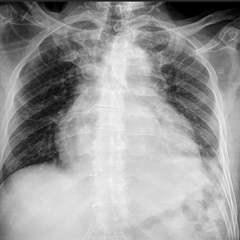
Pericardial effusion has a broad spectrum of clinical presentation, ranging from an incidental finding on imaging to a potentially fatal emergency such as pericardial tamponade, the most severe presentation. The authors present a case of a middle-aged male hospitalized due to shortness of breath. Initial work-up was positive for massive pericardial effusion with haemodynamic compromise. Additional study revealed panhypopituitarism. The acromegalic phenotype was suggestive of acromegaly secondary to pituitary adenoma, which had probably evolved to apoplexy. Hormone replacement was started with clinical improvement. At the 3-year follow-up, there was no evidence of recurrence of pericardial effusion. Panhypopituitarism is a relatively rare entity, but can lead to life-threatening complications such as adrenal crisis, coma and myxoedema-associated cardiac failure. Pericardial effusion is an extremely rare manifestation of secondary hypothyroidism.
|
Cough Syncope, a Rare Presenting Symptom of Chiari I Malformation and Atlanto-Occipital Assimilation
Views: 1492
HTML: 172
PDF: 443
|
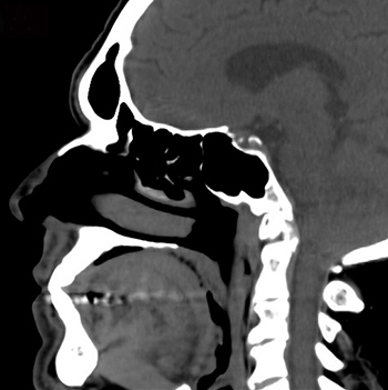
Chiari I malformation and atlanto-occipital assimilation are both fairly uncommon conditions. Symptoms usually present during adolescence or adulthood, typically consisting of headache or neck pain. Cough-associated syncope is an unusual presenting symptom. The diagnosis of this condition in a pulmonology department is even rarer.
We report the case of a 62-year-old male referred to our pulmonology department due to complaints of cough-associated syncope. After several examinations, a pharyngeal CT scan incidentally showed low positioning of the cerebellar tonsils. Cerebral MRI confirmed the diagnosis of Chiari I malformation and atlanto-occipital assimilation and the patient was effectively treated with surgical decompression.
|
Views: 689
HTML: 86
PDF: 369
|
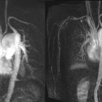
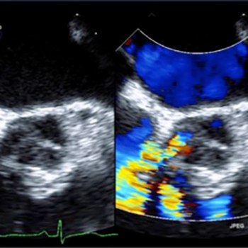
Interrupted aortic arch (IAA) is an extremely rare congenital cyanotic heart disease characterized by complete disruption between the ascending and descending aorta. A patent ductus arteriosus (PDA) or other collateral pathways provide blood flow to the distal descending aorta. Mortality is extremely high at early infancy, particularly after the closure of ductus arteriosus. Survival and presentation in adulthood are extremely rare. Here we illustrate a rare case of type B interrupted aortic arch in an adult who presented with secondary polycythaemia. The blood supply to descending aorta and beyond is almost solely by a patent ductus arteriosus. The case demonstrates the value of multimodality imaging including CT and MRI for diagnosis and treatment planning in these patients.
|
Views: 1581
HTML: 127
PDF: 522
|
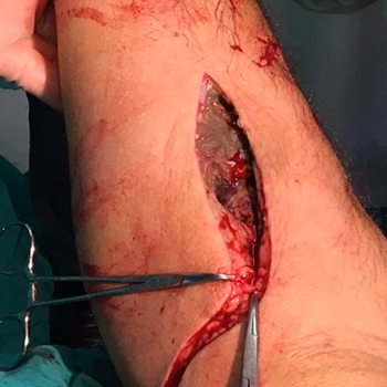
Necrotizing fasciitis is a rare but potentially fatal infection involving the subcutaneous tissue and fascia with the development of necrosis of these structures. Acute compartment syndrome occurs when increased pressure within a closed muscle compartment compromises the circulation and function of the tissues within that space. We report the case of a male patient who was admitted to the intensive care unit for the management of urosepsis due to an acute obstructive pyelonephritis complicated by cardiopulmonary arrest. A radial arterial catheter in the left arm was urgently inserted, under suboptimal aseptic technique. His clinical condition progressively deteriorated, and swelling of the left arm with extension to the forearm with incipient signs of compromised perfusion were observed. The diagnosis of necrotizing fasciitis with acute compartment syndrome was made and an emergency fasciectomy performed. Following this, the patient gradually improved, organ dysfunction resolved, and he was discharged without sequelae.
|
Views: 1219
HTML: 191
PDF: 593
|
Isolated congenital asplenia is a rare condition that mostly manifests in the early years, usually due to fatal systemic infections. In this paper, however, we present a case of a 36-year-old asymptomatic patient who was referred for suspected hyposplenism, with no history of splenectomy. There were no significant changes on physical examination. Blood analysis revealed leukocytosis and thrombocytosis as well as moderate anisopoikilocytosis and red blood cells with Howell–Jolly bodies. No spleen or other malformations were identified on imaging. Individuals with isolated congenital asplenia have an increased susceptibility to invasive infections and sepsis, with rapid clinical decline and a high mortality rate despite treatment.
|
Views: 1195
HTML: 169
PDF: 509
|
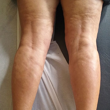
Fasciitis with eosinophilia (FE) is a rare connective tissue disease. Due to its rarity, large-scale studies are lacking, which makes its treatment challenging. Systemic corticosteroids (SCSs) are the cornerstone of treatment; however, additional immunosuppressive drugs (ISDs) are frequently necessary (usually methotrexate). We report 2 patients, for whom an SCS and methotrexate were not a viable long-term option. In the first case, we were unable to taper the SCS dose without symptom relapse, the patient showed only a partial response to methotrexate and presented side effects. The second case never fully responded to the SCS and methotrexate and demonstrated serious SCS adverse effects. Both patients were started on tocilizumab with extremely favourable results, making this drug a potential therapeutic weapon for these patients.
|
Views: 933
HTML: 201
PDF: 607
|
Amyotrophic lateral sclerosis (ALS) rarely presents with hyponatraemia caused by syndrome of inappropriate antidiuretic hormone secretion (SIADH). We present a patient with hyponatraemia of multifactorial aetiology, in whom, after withdrawal of the drugs that contributed to this ionic alteration, SIADH secondary to ALS was confirmed. After initiating treatment with urea, sodium levels were normalized.
|
Views: 1220
HTML: 113
PDF: 522
|
We present a case of an 85-year-old woman diagnosed with uncomplicated pyelonephritis, who was treated with intravenous ceftriaxone. Her chronic medications were phenprocoumon, diltiazem and bisoprolol. During the infectious phase, the patient presented tachycardia – despite high-dose beta-blocker treatment – and developed left acute heart failure, with acute renal failure (pre-renal origin). After introduction of furosemide diuretic therapy, clinical conditions improved and better control of the volemic status and heart rate was achieved. Several days after ceftriaxone and digoxin therapy initiation, worsening multiple non-blanching palpable purpuric lesions with bullae and papules, limited to the lower extremities, were noted. Skin biopsy was performed and a diagnosis of leucocytoclastic vasculitis, with associated panniculitis, was made. Ceftriaxone was discontinued and systemic corticosteroids were introduced, with a clear improvement in the cutaneous condition.
|
Views: 986
HTML: 80
PDF: 395
|
A ruptured sinus of Valsalva aneurysm as a cause of aorto–atrial fistula is very rare. We present the case of a 53-year-old female who presented with symptoms of acute heart failure and suspicion of an aorto–atrial fistula found on a transthoracic echocardiogram, which was confirmed on transesophageal echocardiography. A coronary angiogram showed normal coronary arteries but confirmed the right aorto–atrial fistula on aortogram. She underwent successful surgical repair of the fistula. Her postoperative echocardiogram showed a normal right atrium and right ventricle with no shunt. A ruptured sinus of Valsalva aneurysm is a devastating event and presents as acute heart failure. Prompt diagnosis and surgical repair is necessary to prevent mortality.

|
Views: 1009
HTML: 70
PDF: 383
|
Chronic ingestion of liquorice induces a syndrome with findings similar to those for primary hyperaldosteronism. This is characterized by hypokalaemia, hypertension, metabolic alkalosis and suppression of the renin-aldosterone system.
We describe a 30-year-old woman who, with a plasma potassium level of 1.5 mmol/l, presented with tetraparesis and severe rhabdomyolysis (CK up to 35,460 U/l). She admitted to a daily consumption of nearly 300 g of liquorice sweets during the previous 6 months. This case emphasizes the importance of a detailed anamnesis, which is essential for diagnosis, avoids unnecessary and expensive investigations and reduces the duration of hospitalization.
|
Views: 779
HTML: 102
PDF: 411
|
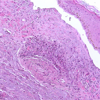
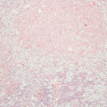
Kikuchi-Fujimoto disease (KFD) is a rare, benign and usually self-limiting disorder that more often affects young women, which is characterized by cervical lymphadenopathy and fever. Clinical presentation may be indistinguishable from other diseases, and its inclusion in the differential diagnosis of lymphoproliferative, infective and autoimmune diseases is essential. An association with systemic lupus erythematosus is acknowledged. We present 2 different cases of 2 young women with KFD; the first case highlights the classic diagnostic features of this rare entity, and the second, the findings when KFD occurs in association with systemic lupus erythematosus.
|
Views: 952
HTML: 75
PDF: 402
|
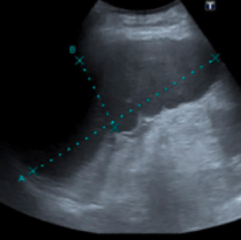
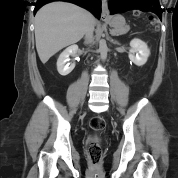
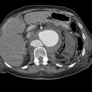
We describe a rare presentation of acute pyelonephritis associated with a ruptured abdominal aortic aneurysm. A 68-year-old female presented to the emergency department with a 3 day history of cystitis. General examination revealed the acute onset of pain in the left flank accompanied by fever and chills. Blood tests revealed leucocytosis 25,400x109L and C-reactive protein 495 mg/L (<6.1), while urinary sediment analysis revealed many leucocytes and gram-negative bacteria. The patient was admitted with acute pyelonephritis. On the third day of admission, the urine culture isolated Escherichia coli sensitive to the antibiotic prescribed; however, the patient clinically deteriorated. A computed tomography scan revealed a ruptured abdominal aortic aneurysm involving the left renal artery. The patient underwent an exploratory laparotomy but uncontrollable haemorrhage led to a fatal outcome.
This case highlights a rare case of acute pyelonephritis associated with a ruptured abdominal aortic aneurysm. A computed tomography scan or abdominal ultrasound should be considered whenever a patient has acute pyelonephritis with a C-reactive protein >400 mg/L in order to exclude complications and other potentially fatal pathologies.
|
Views: 799
HTML: 64
PDF: 357
|
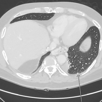
Oesophageal varices are a dilated submucosal venous plexus in the lower third of the oesophagus which result from increased pressure in the portal venous system. The portal system is connected to the systemic circulation in specific locations referred to as sites of portosystemic anastomosis. An increase in portal venous pressure is therefore reflected at these anastomotic sites, causing manifestations such as oesophageal varices, rectal varices, caput medusae and splenorenal shunts. Varices do not cause symptoms until they leak or rupture and this is the main complication which requires prompt treatment. Here, we present a post-liver transplant patient with metastatic hepatocellular carcinoma who had oesophageal varices that fistularized with a left pulmonary vein, thus creating a right-to-left shunt. Right-to-left shunts are usually intracardiac or intrapulmonary in location. The complications of a right-to-left shunt include predominantly hypoxia, cyanosis and, sometimes, paradoxical emboli in the case of intracardiac shunts. This patient had a very uncommon cause of such a shunt caused by a direct fistulous connection.
|
Views: 949
HTML: 61
PDF: 440
|
Intravenous thrombolysis with recombinant tissue plasminogen activator (rtPA) is the established treatment for acute ischemic stroke and has been highly effective in reducing the neurological deficit. Serious adverse events are not uncommon, with hemorrhage being the major complication. We describe the case of a patient with acute ischemic stroke that also presented with vague cardiac symptoms and was treated with rtPA, which was complicated by a hemopericardium causing cardiac tamponade. Pericardiocentesis was promptly performed, which resulted in rapid resolution of the cardiogenic shock. The patient recovered consciousness within a few minutes. A search of the MEDLINE database shows that this is the first report of cardiac tamponade after rtPA thrombolysis occurring in a patient with no history of recent myocardial infarction or aortic dissection.

|
Views: 899
HTML: 324
PDF: 437
|
Postpartum hypoglycemia in non-diabetic women is a rare condition. We report the case of a 34-year-old woman who experienced neuroglycopenia 2 days after delivery. Corresponding to severe hypoglycemia, we found inappropriately elevated insulin and C-peptide levels. Following magnetic resonance imaging a lesion of 10x8 mm was detected in the head of the pancreas. An ultrasound-guided fine needle aspiration of the mass confirmed the diagnostic suspicion of a pancreatic neuroendocrine tumor. Complete surgical enucleation of the insulinoma resulted in immediate and permanent resolution of the hypoglycemia. The postoperative course was uneventful. Histopathological and immunohistochemical analyses were consistent with insulinoma. The diagnostic approach to postpartum hypoglycemia represents a challenge for multidisciplinary teamwork.

|
Views: 3957
HTML: 273
PDF: 1480
|
We describe two elderly patients evaluated at emergency departments for anosmia/dysgeusia in the absence of any other respiratory symptoms prior to or upon admission. In the current epidemiological context, clinical and biological work-up led to a diagnosis of COVID-19 infection. Unfortunately, one of the patients died during hospitalization, but the other recovered and was discharged.
|
Views: 3112
HTML: 446
PDF: 2364
|
COVID-19 (coronavirus disease 19) is an infectious disease caused by coronavirus SARS-CoV-2. Since its detection in China at the end of 2019, the novel coronavirus has rapidly spread throughout the world and has caused an international public health emergency. The most common manifestation is flu-like symptoms. Mild infections usually improve within a few days, but COVID-19 can cause severe pneumonia with acute respiratory distress syndrome and death. Gastrointestinal symptoms are less common but possible and more difficult to recognize as part of a COVID-19 syndrome. In line with the current opinion of the WHO, we strongly believe that preventive measures and early diagnosis of COVID-19 are crucial to interrupt virus spread and avoid local outbreaks. We report the cases of COVID-19 patients admitted to our Emergency Department who complained of gastrointestinal symptoms at admission.
|
Views: 1112
HTML: 274
PDF: 674
|
Amiodarone is an antiarrhythmic drug, in use from the 1960s, which acts on potassium transport in myocytes, causing a lengthening of the action potential and refractory period. Even though it is broadly prescribed, its use is limited by a relatively high occurrence of adverse reactions such as lung, thyroid or hepatic disease, skin changes and so on. The authors report a case of a female patient who was admitted due to chest pain. Due to the bluish skin pigmentation, other causes of amiodarone toxicity were investigated, and hyperthyroidism was detected. After amiodarone discontinuation and specific therapy, thyroid function returned to normal.
| 2.1 = | 1.762 Cit. to date |
| 842 Docs. to date |


Publisher
Official Journal of the
European Federation of Internal Medicine
www.efim.org
Publisher: SMC media Srl
Via Giovenale, 7 - 20136 Milan - Italy
P.IVA 07626490960
info@ejcrim.com
www.ejcrim.com - ISSN: 2284-2594 - © EFIM 2014-2024, Published by SMC Media srl, Italy - Privacy policy

