EJCRIM 2023 CiteScore
| 2.1 = | 1.762 Cit. to date |
| 842 Docs. to date |
Last updated on 05 May, 2024
Updated monthly
Updated monthly
Powered by

|
Views: 1225
HTML: 785
PDF: 628
|
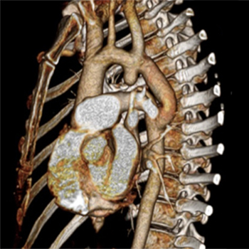
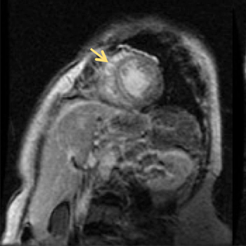
Interrupted aortic arch (IAA) is a rare congenital abnormality with only a few cases reported in adults. It is defined as complete loss of continuity between the ascending and descending portions of the aorta, and is usually associated with other cardiac defects. The diagnosis in adults should be suspected in the presence of refractory hypertension, a careful physical examination being crucial to early diagnosis. Magnetic resonance angiography (MRA) techniques can accurately characterize cardiovascular anatomy, and also provide information regarding heart chamber and valve function.
|
Views: 1169
HTML: 195
PDF: 430
Acute paraplegia with cognitive alterations after bilateral infarcts in cerebral small vessel disease: 0
|
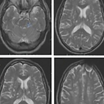
Cerebral small vessel disease (SVD) affects the small arteries, arterioles, venules and capillaries in the brain and can be identified clinically and/or radiologically. We describe the case of a 71-year-old man with sporadic cerebral SVD who presented with acute paraplegia with urinary incontinence and recent cognitive impairment that developed after the occurrence of ischaemic lesions.
|
Views: 1216
HTML: 344
PDF: 581
Syphilis tables and images: 0
|
Syphilis is one of the oldest described infectious diseases in the world and is caused by the spirochete bacterium Treponema pallidum[1]. Although now a rare disease, incidence is increasing with the number of diagnoses of the disease rising in England from 1688 to 2713 between 2003 and 2012 (a 61% increase)[2]. Major outbreaks of syphilis have been documented in London, Manchester, Dublin, and Brighton particularly among men who have sex with men (MSM)[3]. Diagnosis remains difficult on account of multi-system symptoms, duration of the condition, and social stigma.
|
Views: 1209
HTML: 358
PDF: 612
Senile Systemic Amyloidosis - an underdiagnosed disease: 0
|
Senile systemic amyloidosis is caused by a non-mutated form of transthyretin with the heart being the major organ involved. This infiltrative cardiomyopathy usually presents as slowly progressive heart failure.
An 82-year-old female patient was admitted for newly diagnosed heart failure. A year later she presented with decompensated heart failure and syncope. Inpatient work-up showed persistently elevated troponin and N-terminal-pro BNP levels, rapid progression to severe left ventricular concentric hypertrophy, and sinus pauses on the Holter. Cardiac MRI revealed diffuse late gadolinium enhancement in the left ventricle. The demonstration of amyloid protein with the clinical findings and complementary investigations allowed for the diagnosis of senile systemic amyloidosis.
|
Views: 2143
HTML: 300
PDF: 518
|
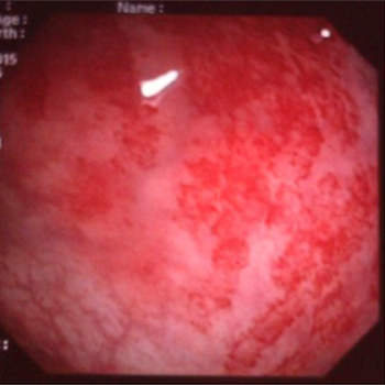
Henoch-Schönlein purpura is an IgA-mediated immune vasculitis which is characterized by purpuric lesions and osteoarticular, intestinal and sometimes renal manifestations. The histopathological substrate of this entity is leucocytoclastic vasculitis (LCV) with IgA deposits seen on immunohistochemistry. We here report the case of a 27-year-old woman with abdominal pain and cutaneous purpura. Upper and lower endoscopic exploration showed purpuric lesions in the rectum but not in the stomach. Skin biopsy revealed LCV. IgA deposits were seen only in gastric mucosa. The patient was treated with corticoids which led to improvement of both the cutaneous and digestive symptoms. This case suggests that gastrointestinal biopsies of both normal and abnormal mucosa should be taken in Henoch-Schönlein purpura, especially in patients with atypical forms.
|
Views: 882
HTML: 209
PDF: 428
Figures: 0
Parasternal long axis view showing the biventricular HCM: 0
Parasternal short axis view showing the biventricular HCM: 0
Apical four chamber view showing the biventricular HCM: 0
|
Light-chain (AL) amyloidosis is the most common type of amyloidosis; cardiac involvement is rare but has a poor prognosis. Biventricular hypertrophic cardiomyopathy is an exceptional finding in amyloidosis and its association with obstructive right ventricular gradient is even rarer. We report the case of a male patient with biventricular hypertrophy suggesting amyloidosis, with an obstructive gradient in the right ventricle.
| 2.1 = | 1.762 Cit. to date |
| 842 Docs. to date |


Publisher
Official Journal of the
European Federation of Internal Medicine
www.efim.org
Publisher: SMC media Srl
Via Giovenale, 7 - 20136 Milan - Italy
P.IVA 07626490960
info@ejcrim.com
www.ejcrim.com - ISSN: 2284-2594 - © EFIM 2014-2024, Published by SMC Media srl, Italy - Privacy policy

