

EJCRIM 2023 CiteScore
| 2.1 = | 1.762 Cit. to date |
| 842 Docs. to date |
Last updated on 05 May, 2024
Updated monthly
Updated monthly
Powered by

|
Views: 318
PDF: 241
HTML: 29
|
Introduction: There are very few documented cases of Escherichia coli endocarditis with cardiac abscesses in the literature. Here we describe a case presentation with diagnostic challenges and a multidisciplinary approach to management.
Case description: This is a rare presentation of E. coli endocarditis in a patient with a prosthetic aortic valve. Initial tests were inconclusive and further investigation with transoesophageal echocardiography was required to make the diagnosis. Despite initial improvement, the patient deteriorated and ultimately died of complications related to the presentation.
Discussion/conclusion: E. coli is a rare causative organism for endocarditis, which can itself be difficult to diagnose. A multidisciplinary approach to investigation and treatment is required when infective endocarditis is suspected. Transoesophageal echocardiography may be required to diagnose endocarditis when there is a strong clinical suspicion and risk factors present.
|
Views: 292
HTML: 46
PDF: 199
|
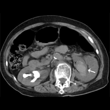
Subepithelial haemorrhage of the renal pelvis is a rare cause of haematuria and can be diagnosed based on radiographic findings. This haemorrhage often appears as a non-enhancing hyperdense mass in the renal pelvis on computed tomography, which sometimes results in unnecessary nephrectomy because it can mimic renal neoplasms. It can be managed conservatively, and its prognosis is generally benign. We report a case of renal pelvic haemorrhage complicating emphysematous pyelonephritis that needed emergent nephrectomy. Our case highlights the importance of careful observation for complications of urinary tract infection, although complications are rare.
|
Views: 371
HTML: 50
PDF: 287
|
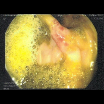
Introduction: Hereditary haemorrhagic telangiectasia (HHT) is a rare multi-organ vascular disease. It is characterised by mucocutaneous telangiectasia, epistaxis, and arteriovenous malformations. Some 70% of patients with HHT are thought to have issues with gastrointestinal (GI) bleeding. Traditional management of GI bleeding in HHT includes monitoring for iron deficiency anaemia, iron replacement, antifibrinolytic therapy and control of identifiable bleeding sites with argon photocoagulation during gastrointestinal endoscopy. Blood transfusion may also be required.
Case description: Our case describes a man in his 40s with confirmed HHT, with transfusion-dependent anaemia secondary to GI bleeding. He was commenced on fortnightly bevacizumab (5 mg/kg) for 12 weeks in an attempt to reduce his blood transfusion requirement and manage his anaemia. In the months prior to starting bevacizumab, our patient’s transfusion requirement ranged from 3–5 units of packed red cells per month to maintain an Hb >8 g/dl. He had a marked improvement in his symptoms within the first month of treatment and did not require any further blood transfusion during the three months of treatment. He was given one further IV iron infusion in the final month of his 3-month bevacizumab treatment and did not experience any adverse side effects from bevacizumab.
Discussion: HHT results from alterations to genes which encode proteins involved in blood vessel formation. This provides the rationale for using anti VEGF drugs such as bevacizumab. Current evidence for this treatment approach is limited.
Conclusion: Bevacizumab can be an effective treatment option in patients with HHT refractory to traditional management.

|
Views: 658
HTML: 74
PDF: 519
|
Sodium-glucose cotransporter-2 (iSGLT2) inhibitors, which include dapagliflozin, canagliflozin and empagliflozin, are a class of drugs initially used in the oral treatment of diabetes, heart failure and renal failure. They target the reabsorption of glucose in the kidney. Although they bring benefit to patients with these conditions and in general produce few adverse effects, in some cases, iSGLT2 can cause serious adverse effects such as metabolic acidosis, and fungal or bacterial urinary infections. Oncology patients, who in general have a weak immune system and are usually treated with chemotherapy and/or immunotherapy, are more susceptible to this type of adverse events than other patients. For this reason, it is necessary to adequately select the patients eligible to receive this type of drug and evaluate the potential benefits for them. In this series of five cases, we present two cases of metabolic acidosis, two cases of bacterial urinary sepsis, and one case of fungal urinary sepsis that occurred in patients admitted to the Medical Oncology Department of the University Hospital of Salamanca in 2023.

|
Views: 363
HTML: 28
PDF: 281
|
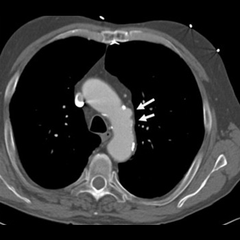
Immunoglobulin G4-related disease (IgG4-RD) is an autoimmune disease characterized by elevated serum IgG4 levels. It has the potential to affect multiple organs. Despite the diverse manifestations of IgG4-RD, the association with coronary artery disease (CAD) remains poorly understood due to limited evidence. We report the case of a 52-year-old male patient who exhibited typical angina upon exertion, accompanied by elevated serum IgG4 levels. Coronary computed tomographic angiography (CCTA) revealed the presence of pseudotumor formations surrounding and aneurysm changes affecting all coronary arteries, consistent with IgG4-RD. The patient was treated with prednisolone and azathioprine, with the possibility of additional rituximab therapy if symptomatology failed to improve. This case sheds light on the rare occurrence of IgG4-RD with coronary artery involvement and underscores the importance of recognizing this unique clinical entity for appropriate management and further research.
|
Views: 257
HTML: 15
PDF: 223
|
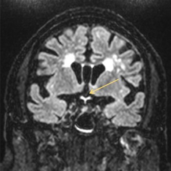
Acute bilateral blindness is an emergent condition that may signal life-threatening disease. The approach focuses on identification of life-threatening disease processes, while differentiating between ocular, psychogenic and neurologic aetiologies. We present the case of an 88-year-old man with multiple cardiovascular risk factors and bilateral chronic glaucoma and cataracts. He presented to the emergency department with sudden onset bilateral loss of visual acuity with no other relevant changes on physical examination, including other neurologic deficits. Ophthalmologic observation showed no sign of acute disease.
Contrast orbit and cranioencephalic CT was unremarkable, so the patient underwent an orbit and cranioencephalic MRI that showed changes in fluid-attenuated inversion recovery (FLAIR) sequences and diffusion restriction involving the optic chiasm and the initial segment of the optic radiations bilaterally. Optic chiasm strokes are rare, owing to the rich supply of collateral circulation. The most frequent presentation is bitemporal hemianopsia but rarer presentations are described. Bilateral loss of visual acuity is very rare and infarction of the whole optic chiasm is unusual.

|
Views: 288
HTML: 24
PDF: 254
|
Background: Epididymitis is a common cause of scrotal pain in adults, with coliform bacteria being the most common isolated organisms in patients older than 35.
Case presentation: A 51-year-old healthy patient presented with scrotal pain and swelling, and was found to have epididymo-orchitis and bacteraemia caused by Haemophilus influenzae, which has not previously been reported as a cause of epididymo-orchitis and bacteraemia in immunocompetent patients.
Discussion: Diagnostic studies can help confirm the diagnosis and detect the causative pathogen. In all suspected cases, a urinalysis, urine culture and a urine or urethral swab for nucleic acid amplification tests (NAATs) for Neisseria gonorrhoeae and Chlamydia trachomatis should be performed. Colour Doppler ultrasonography often shows an enlarged thickened epididymis with increased Doppler wave pulsation in epididymitis. H. influenzae are pleomorphic gram-negative rods that commonly colonise the human respiratory tract and are associated with a number of clinical conditions. H. influenzae has been reported as a cause of epididymo-orchitis in prepubertal boys, and in few cases were associated with positive blood cultures. In adults, H. influenzae has been isolated before from urine samples or urethral swabs in patients with epididymitis or epididymo-orchitis.
Conclusion: This case highlights the possibility of H. influenzae causing epididymo-orchitis and bacteraemia in immunocompetent patients. Healthcare providers should consider H. influenzae in the differential diagnosis of epididymitis and epididymo-orchitis in both immunocompetent and immunocompromised patients.
|
Views: 362
HTML: 43
PDF: 237
|
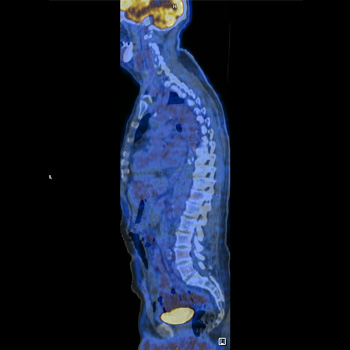
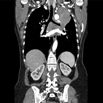
Q fever is a zoonotic infection caused by the pathogen Coxiella burnetii, and patients can present with a wide spectrum of clinical manifestations, depending on whether it is an acute or a chronic infection.
We present the case of a 61-year-old male with fatigue, posterior thoracalgia, intermittent fever, night sweats and weight loss for a month. After an extensive workup, he was diagnosed with acute Q fever with large-vessel vasculitis. The FDG-PET/CT scan suggested an active vasculitis specifically in the thoracic aorta, proximal abdominal aorta, subclavian and carotid vessels, suggesting an immunologic response to acute Q fever infection, barely reported worldwide.
|
Views: 361
HTML: 28
PDF: 289
|
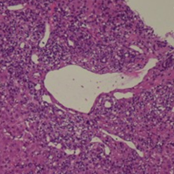
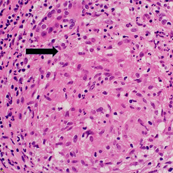
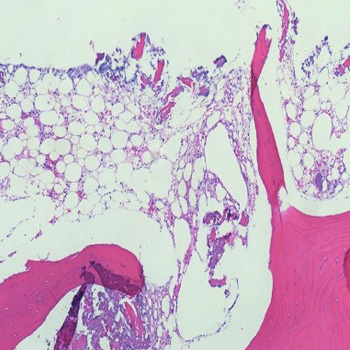
Introduction: Recently, medical interest has been growing in SARS-CoV-2 infection and its multiorgan involvement, including the liver. Up until now, a few reports have described autoimmune hepatitis (AIH) triggered by SARS-CoV-2 infection, but no data are available about the specific liver inflammatory infiltrate and cluster of differentiation. We report a case of AIH triggered by SARS-CoV-2 infection, with a particular focus on its histological and mainly immunohistochemical features.
Case description: A 60-year-old man, with a history of paucisymptomatic SARS-CoV-2 infection that occurred one month earlier, was admitted for alterations of hepatocellular necrosis and cholestasis indexes. He completed vaccination for SARS-CoV-2 a year earlier. The serologies for hepatotropic viruses were negative. The anti- smooth muscle antibodies (ASMA) and antinuclear antibodies (ANA) results were positive. Anti-liver kidney microsome (anti-LKM) antibodies and antimitochondrial (AMA) were negative. By liver biopsy, haematoxylin-eosin staining highlighted severe portal inflammation with a rich CD38+ plasma cell component, while immunohistochemical staining showed low cell CD4+ count and prevalence of CD8+ and CD3+. After biopsy, the patient started an immunosuppressant regimen, with benefit.
Discussion: We can conclude that the patient developed a type 1 AIH triggered by SARS-CoV-2 infection. The presence of CD8 T-cells at immunohistochemical examination suggests different mechanisms from classic AIH. Similar cases are described after AIH triggered by SARS-CoV-2 vaccination.
Conclusion: The AIH after SARS-CoV-2 infection developed by the patient showed a histological picture similar to a classic AIH for the abundant presence of plasma cells, and immunohistochemical features similar to those described after SARS-CoV-2-vaccination.
|
Views: 278
HTML: 38
PDF: 218
|
This report presents the clinical details and management of a 58-year-old Caucasian male with pericardial effusion and cardiac tamponade following outpatient inferior vena cava (IVC) filter removal. The patient was unresponsive and experienced cardiac arrest minutes after the procedure, requiring cardiopulmonary resuscitation. After return of spontaneous circulation he displayed somnolence, confusion and chest discomfort. Investigations revealed a large pericardial effusion, and an echocardiography confirmed cardiac tamponade. Prompt intervention involved pericardiocentesis, resulting in haemodynamic stabilisation and reduction in effusion size. The patient responded favourably with treatment. Differential diagnoses were considered and treatment options were discussed, highlighting the importance of timely recognition and appropriate intervention in managing pericardial effusion and cardiac tamponade. This report adds to the limited literature on pericardial effusion and cardiac tamponade following a scheduled outpatient IVC filter removal, emphasising the unique clinical presentation and successful management of this rare phenomenon.
|
Views: 586
HTML: 23
PDF: 364
|
Introduction: Most lung cancers are diagnosed at an advanced stage. Common metastatic sites include the brain, bone, liver and adrenal glands. Ocular metastases, however, are extremely rare. We present a case of advanced lung adenocarcinoma presenting exclusively with photopsias attributable to retinal metastases.
Case description: We describe a woman in her fifties, a lifetime non-smoker with an unremarkable medical and family history, who presented to the emergency department with photopsias for a week. Ophthalmology evaluation revealed decreased visual acuity bilaterally, and a fundus examination disclosed lesions suggestive of bilateral retinal metastases. A comprehensive evaluation diagnosed a stage IVb lung adenocarcinoma with exon 19 mutation on epidermal growth factor receptor gene. Subsequently, she developed complaints of headaches and dizziness. She received frontline osimertinib 80 mg daily, preceded by upfront whole-brain radiation therapy with partial orbital inclusion for symptomatic ocular and brain metastases. After ten radiation therapy sessions, her complaints were resolved and an ophthalmology revaluation revealed improvement in visual acuity and resolution of photopsia complaints. The patient is currently on osimertinib and preserves an ECOG score of 0.
Conclusion: Retinal metastases usually indicate advanced disease, so presenting with isolated ocular symptoms is exceedingly rare. Especially in cases of uncommon metastases, a multidisciplinary approach is fundamental for a prompt diagnosis and timely treatment, impacting prognosis and quality of life.

|
Views: 2629
HTML: 454
PDF: 538
|
Introduction: High blood concentrations of vitamin B12 are often caused by over-supplementation. However, there are instances in which augmented vitamin B12 levels are seen in the absence of supplements. Macro-vitamin B12 is an underrated cause of supra-physiological cobalamin plasma levels.
Case description: A 70-year-old man was referred to an ambulatory internal medicine centre because of high vitamin B12 levels yet he denied taking supplements. An X-ray showed a tumour in the right upper lobe of the lung, which triggered further examinations. An MRI scan of the brain came back normal as well as a CT scan of the abdomen, and colonoscopy. The pulmonologist requested a PET-CT scan, which showed an isolated 18-FDG uptake in the area of the lung mass that was detected earlier. The patient underwent surgery with adjuvant cis-platinum and gemcitabine and is still making good progress. The vitamin B12 levels persisted after successful treatment of lung adenocarcinoma; determination of vitamin B12 after PEG (polyethylene glycol) precipitation showed normal concentrations.
Discussion: A high vitamin B12 plasma concentration in the absence of vitamin supplementation can be a daunting diagnostic problem for the internist, as there are several possible underlying causes. In this case the diagnosis of lung carcinoma was made, the patient was treated appropriately, yet this pathology had no correlation with the cobalamin levels.
Conclusion: A high vitamin B12 concentration can be the impetus of thorough medical inquiries. Internists should be careful not to forget macro-vitamin B12 as a possible source of falsely elevated vitamin B12 values.
|
Views: 444
HTML: 35
PDF: 283
|
Corynebacterium spp. are Gram-positive bacteria, and recent studies have proposed a potential link between granulomatous mastitis and Corynebacterium kroppenstedtii infections, posing a challenge in selecting appropriate antibiotics, particularly in pregnant women. A young pregnant woman presented with a palpable lump in her left breast. Subsequent assessment revealed the presence of necrotising granulomatous mastitis attributed to C. kroppenstedtii. Initially treated with amoxicillin/clavulanate, the patient showed no improvement. Consequently, clindamycin was administered based on culture and sensitivity results, which resulted in a favourable response with no recurrence of symptoms. This report aims to emphasise the efficacy of clindamycin as a treatment option for granulomatous mastitis caused by C. kroppenstedtii.
|
Views: 273
HTML: 50
PDF: 252
|
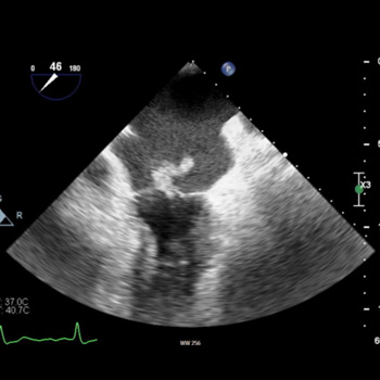
Background: Aortic dissection is a critical condition often presenting with acute, severe chest pain and haemodynamic instability. Early diagnosis is essential to mitigate the high mortality risk. Imaging modalities play a pivotal role in diagnosing aortic conditions, but determining the appropriate method can be challenging.
Case description: We report an asymptomatic 55-year-old female undergoing transthoracic echocardiography (TTE) for mitral and aortic valve regurgitation surveillance. Incidentally, a suspicious flow jet at the distal aortic arch was discovered, raising concerns of aortic dissection. A subsequent CT angiogram (CTA) identified this as an aortic ductus diverticulum at the aortic isthmus, not a dissection.
Discussion: Aortic dissection has a high initial 48-hour mortality, with even surgically managed cases exhibiting a 26% risk. Imaging tools such as a transoesophageal echocardiogram (TEE), CT and MRI scans are instrumental for diagnosis, with their applicability depending on the patient’s clinical situation. The aortic ductus diverticulum, a developmental outpouching, often mimics other aortic pathologies, emphasising the importance of accurate imaging interpretation.
Conclusion: Aortic ductus diverticulum presents diagnostic challenges due to its resemblance to other aortic conditions. Advancements in imaging modalities improve diagnostic accuracy, but awareness and careful interpretation are paramount to ensure timely and appropriate patient care.
|
Views: 391
HTML: 57
PDF: 279
|
Onychomycosis (OM), a widespread fungus that affects the toenails and/or fingernails, causes a large amount of morbidity and is very frequent in the general population. The best treatment is systemic antifungals. Terbinafine is a potent antifungal drug that works by targeting the keratin and lipids found in fungi. In the United States, the prevalence of this nail ailment ranges from 2% to 14%; it is 5.5% globally. Here, we describe a case of aplastic anaemia linked to oral terbinafine use. Clinicians should be aware of this rare adverse effect and early discontinuation of the treatment is required to prevent significant morbidity and mortality.

|
Views: 391
PDF: 343
HTML: 38
|
Mucopolysaccharidosis type IVA (MPS-IVA) is a rare lysosomal storage disease caused by N-acetylglucosamine-6-sulfate-sulfatase enzyme deficiency. MPS-IVA patients show severe extra-skeletal and skeletal manifestations, featured by bone pain and deformities, frailty fractures and early onset osteoporosis. The enzyme replacement therapy (ERT) with elosulfase alpha stabilizes the MPS-IVA extra-skeletal manifestations but does not significantly improve MPS-IVA skeletal manifestations. We administered an integrated therapy to an MPS-IVA 41-year-old male patient, composed of zoledronic acid, cholecalciferol and a normocalcemic (calcium intake >1 g/day), hyposodic (sodium intake <5 g/day), and normocaloric diet (bone-diet), other than ERT. During the six-year follow-up, the patient did not develop any adverse events, obtaining an improvement of bone mineral density and quality of life. Given our results, we propose this integrated treatment (i.e. ERT, zoledronic acid, cholecalciferol, and bone diet) in the management of MPS-IVA adult patients.
| 2.1 = | 1.762 Cit. to date |
| 842 Docs. to date |


Publisher
Official Journal of the
European Federation of Internal Medicine
www.efim.org
Publisher: SMC media Srl
Via Giovenale, 7 - 20136 Milan - Italy
P.IVA 07626490960
info@ejcrim.com
www.ejcrim.com - ISSN: 2284-2594 - © EFIM 2014-2024, Published by SMC Media srl, Italy - Privacy policy

