EJCRIM 2023 CiteScore
| 2.1 = | 1.762 Cit. to date |
| 842 Docs. to date |
Last updated on 05 May, 2024
Updated monthly
Updated monthly
Powered by


|
Views: 364
HTML: 45
PDF: 404
|
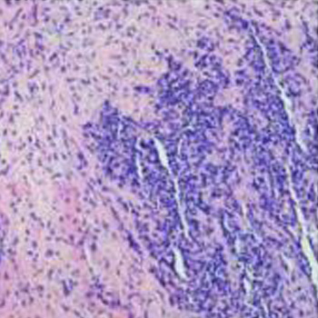
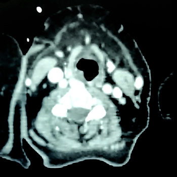
Introduction: Rhabdomyosarcoma is a high-grade malignant neoplasm with skeletal muscle differentiation; a common soft tissue sarcoma in children but considered one of the rarest in adults.
Case description: We report a case of 35-year-old male with a chronic productive cough and haemoptysis for five days. A CT scan of the nasopharynx revealed a blocked left maxillary and ethmoid sinus with bone destruction. These findings raised a suspicion of a tumour, and trans-nasal endoscopic sinus surgery was performed.
Discussion: Nasal rhabdomyosarcoma is a rare adult malignant tumour. Most patients have lymphatic metastasis or skull base tumour infiltration at the time of the initial diagnosis and treatment, which poses a challenge to the diagnosis and management.
Conclusion: Nasal acinar rhabdomyosarcoma, one of the histopathological types of rhabdomyosarcoma, has rapid disease progression and high mortality. Therefore, the treatment of rhabdomyosarcoma requires a combination of surgery, chemotherapy, radiotherapy, and immunotherapy to work together to achieve the best care for the patient.
|
Views: 397
HTML: 59
PDF: 353
|
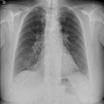
Dasatinib is a tyrosine kinase inhibitor used for treatment of some specific types of leukaemia. The development of pleural effusion is a known adverse effect of dasatinib and chylothorax is exceptional. No case has been reported beyond 5 years of treatment and extensive search for an alternative diagnosis is currently suggested in such scenario. The underlying mechanism is not currently clear. We describe a woman on dasatinib treatment for more than 10 years who developed chylothorax. Drug withdrawal resolved the chylous pleural effusion. We were able to find 14 additional cases of dasatinib-related chylothorax reported up until now.
|
Views: 394
HTML: 67
PDF: 341
|
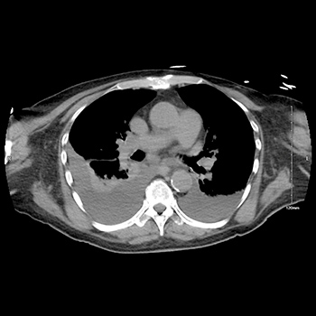
Tracheoesophageal prosthesis (TEP) is an artificial connection between the trachea and esophagus allowing air into the upper esophagus from the trachea thereby vibrating it. TEPs give patients who lose their vocal cords to laryngectomies a tracheoesophageal voice. A potential complication of this is silent aspiration of gastric content. We present a case of a 69-year-old female with a TEP placed after a laryngectomy for laryngeal cancer who presented to the hospital with shortness of breath and hypoxia. She was initially treated for a presumed diagnosis of chronic obstructive pulmonary disease (COPD) and congestive heart failure (CHF) exacerbations but continued to be hypoxic despite aggressive medical management. Further evaluation revealed silent aspirations as a consequence of TEP malfunction. Through our case report we urge clinicians to consider this differential diagnosis, as the clinical presentation of silent aspiration among patients with a TEP can be easily mistaken for a COPD exacerbation. A large number of patients with TEPs are smokers with underlying COPD.
|
Views: 575
HTML: 75
PDF: 494
|
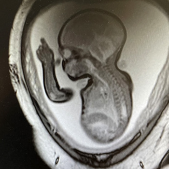
Introduction: Pulmonary embolism (PE) is a rare, severe complication in pregnancy, in which case thrombolysis can be lifesaving but has risks. We aim to highlight actions specific to pregnant women.
Case Description: A 24-week pregnant woman developed shortness of breath and experienced sudden cardiac arrest. Cardiopulmonary resuscitation (CPR) was begun immediately in the ambulance and a perimortem caesarean section was performed upon arrival at hospital, but the new-born died. After 55 minutes of CPR, bedside echocardiography revealed right ventricular strain and thrombolysis was given. The uterus was bandaged to minimize blood loss. After massive transfusions and correction of haemostasis, a hysterectomy was performed due to inability of the uterus to contract. After 3 weeks, the patient was discharged in good health and placed on continuous anticoagulant treatment with warfarin.
Discussion: Approximately 3% of all out-of-hospital cardiac arrest cases are due to PE. Among the few patients who survive at the scene, thrombolysis can be lifesaving and should be considered in pregnant women with unstable PE. Prompt collaborative diagnostic work-up in the emergency room is necessary. In a pregnant woman with cardiac arrest, a perimortem caesarean section improves the chances of both maternal and fetal survival.
Conclusion: Thrombolysis should be considered for patients with PE in pregnancy with the same indications as in a non-pregnant woman. In case of survival, there is profuse bleeding with need for massive transfusions and haemostasis correction. Despite being in very poor condition, the above patient survived and was fully restored to health.
|
Views: 934
HTML: 841
PDF: 504
|
The alkaloid derivatives of Mitragyna speciosa, commonly known as kratom, pose a threat to society due to its potential for abuse, adverse reactions and tendency to be used as self-medication for opioid withdrawal, pain and mood disorders. A number of deaths have been reported along with complications such as respiratory depression, cardiopulmonary arrest, torsade de pointes and seizures. Its various effects and potential are yet to be fully studied. We describe the case of a healthy young male who presented with progressive respiratory failure requiring mechanical ventilation. Imaging revealed multifocal lung infiltrates while extensive infectious and cardiac work-up was negative. Based on the clinical course, a diagnosis of acute respiratory distress syndrome (ARDS) caused by kratom was made. The patient showed gradual clinical improvement and was weaned off supplemental oxygen. This case highlights yet another adverse reaction to kratom and the growing threat posed by its use.
|
Views: 380
HTML: 114
PDF: 383
|
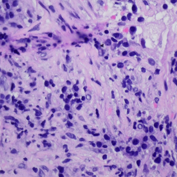
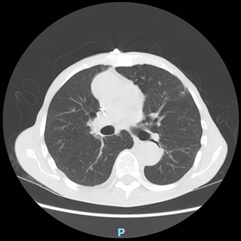
Introduction: Inflammatory myofibroblastic tumours are rare neoplasms which most commonly affect children and young adults. With an intermediate malignant potential, they are typically detected in the abdomen, lung, mediastinum, head and neck, gastrointestinal tract, and genitourinary tract.
Case description: We describe the case of a 33-year-old postpartum woman incidentally diagnosed with a pulmonary inflammatory myofibroblastic tumour following complaints of poorly controlled hypertension a week after caesarean section. She was ALK-negative and received an ALK inhibitor with complete resolution of the lesion. A ROS1–TFG fusion confirmed the diagnosis of an inflammatory myofibroblastic tumour after CT-guided fine needle aspiration.
Discussion: This case highlights an uncommon presentation posing a diagnostic and therapeutic challenge and the potential treatment option of crizotinib.
|
Views: 359
HTML: 67
PDF: 461
|
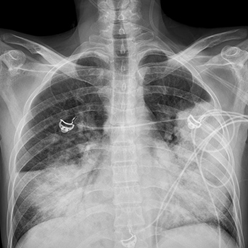
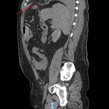
Rib fractures are an infrequent consequence of severe cough. In some patients, undetected rib fractures can lead to life-threatening outcomes. The case of a 73-year-old man who presented with shortness of breath and a worsening dry cough from a SARS-CoV-2 infection for 4 weeks is described. In the emergency department, he was found to be hypoxic and hypotensive. Imaging studies revealed a large right pleural effusion, multiple rib fractures, and right-sided herniation of the colon into the chest. He was admitted to the cardiothoracic intensive care unit where he underwent a flexible bronchoscopy, right video-assisted thoracoscopic surgery, evacuation of a haemothorax, complete decortication, and repair of a diaphragmatic hernia. This case is an unusual presentation of an amalgamation of rare complications resulting from an unrelenting, poorly controlled SARS-CoV-2 infection cough that prompted rapid recognition and swift action.

|
Views: 390
HTML: 42
PDF: 242
|
Endobronchial ultrasound-guided transbronchial needle aspiration (EBUS-TBNA) is a diagnostic tool used to investigate mediastinal lesions. It has a good safety profile, but there are rare accounts of potentially deadly complications. The present article describes one such complication: pericardial empyema.
A 70-year-old man underwent EBUS-TBNA for the differential diagnosis of a pulmonary mass with multiple mediastinal adenopathies. Two weeks after the procedure he developed chest pain, shortness of breath and fever, with rapid progression to hypotension, tachycardia and low peripheral saturation. He was diagnosed with purulent pericarditis with cardiac tamponade. Pericardial drainage and antibiotic therapy were employed with successful recovery from obstructive disease and septic shock.
|
Views: 1135
HTML: 325
PDF: 478
|
Organizing pneumonia (OP) is a form of interstitial lung disease that develops in response to acute lung injury. SARS-CoV-2 causes a wide range of lung and extrapulmonary disease, but there are few data suggesting an association between COVID-19 and OP. We describe a patient with COVID-19 pneumonia who developed severe progressive OP with significant morbidity.
|
Views: 369
PDF: 297
HTML: 47
|
Rosai-Dorfman disease (RDD) is an uncommon lymphoproliferative disorder; RDD with oropharyngeal involvement is extremely rare, especially in adults. A 65-year-old woman with a complaint of progressive dyspnoea since 2016 presented with laryngeal involvement of RDD. A laryngoscopy examination revealed two solid, polypoid masses in the subglottic region, and a laryngeal biopsy concluded chronic in ammation without signs of malignancy. A second biopsy of axillary lymph nodes was performed, supporting the diagnosis of histiocytosis. The patient was treated with corticosteroids and then lost to follow-up. In 2019, she suffered from dyspnoea and a hoarse voice. Laryngoscopy examination showed a polypoid lesion causing airway obstruction at 70% and thickening of the lateral wall of the cavum. Physical examination found left axillary and submandibular adenopathy, and computed tomography revealed thickening of the supraglottic larynx narrowing the laryngeal pathway. Lymphadenectomy with immunohistochemical analysis revealed typical protein positive S-100 histiocytes and emperipolesis. The patient was treated with high doses of corticosteroids for six weeks then these were progressively decreased. The outcome was favourable; the laryngeal lesion disappeared after two weeks of treatment.
| 2.1 = | 1.762 Cit. to date |
| 842 Docs. to date |


Publisher
Official Journal of the
European Federation of Internal Medicine
www.efim.org
Publisher: SMC media Srl
Via Giovenale, 7 - 20136 Milan - Italy
P.IVA 07626490960
info@ejcrim.com
www.ejcrim.com - ISSN: 2284-2594 - © EFIM 2014-2024, Published by SMC Media srl, Italy - Privacy policy

