EJCRIM 2023 CiteScore
| 2.1 = | 1.762 Cit. to date |
| 842 Docs. to date |
Last updated on 05 May, 2024
Updated monthly
Updated monthly
Powered by


|
Views: 3594
HTML: 565
PDF: 562
|
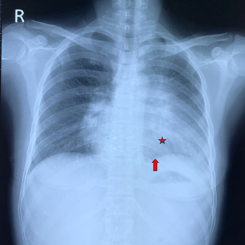
Introduction: High blood concentrations of vitamin B12 are often caused by over-supplementation. However, there are instances in which augmented vitamin B12 levels are seen in the absence of supplements. Macro-vitamin B12 is an underrated cause of supra-physiological cobalamin plasma levels.
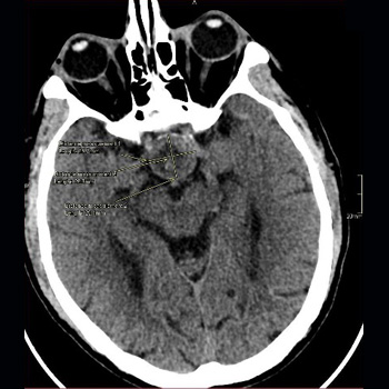
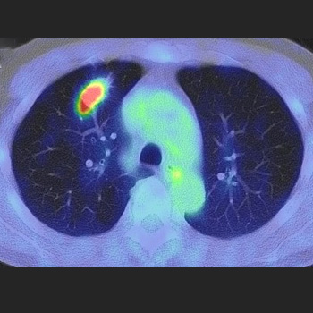
Case description: A 70-year-old man was referred to an ambulatory internal medicine centre because of high vitamin B12 levels yet he denied taking supplements. An X-ray showed a tumour in the right upper lobe of the lung, which triggered further examinations. An MRI scan of the brain came back normal as well as a CT scan of the abdomen, and colonoscopy. The pulmonologist requested a PET-CT scan, which showed an isolated 18-FDG uptake in the area of the lung mass that was detected earlier. The patient underwent surgery with adjuvant cis-platinum and gemcitabine and is still making good progress. The vitamin B12 levels persisted after successful treatment of lung adenocarcinoma; determination of vitamin B12 after PEG (polyethylene glycol) precipitation showed normal concentrations.
Discussion: A high vitamin B12 plasma concentration in the absence of vitamin supplementation can be a daunting diagnostic problem for the internist, as there are several possible underlying causes. In this case the diagnosis of lung carcinoma was made, the patient was treated appropriately, yet this pathology had no correlation with the cobalamin levels.
Conclusion: A high vitamin B12 concentration can be the impetus of thorough medical inquiries. Internists should be careful not to forget macro-vitamin B12 as a possible source of falsely elevated vitamin B12 values.
|
Views: 515
HTML: 57
PDF: 282
|
Primary pulmonary T-cell lymphoma (PPTL) is a rare disease. Diagnosing PPTL is challenging due to non-specific clinical symptoms and imaging. A 32-year-old female presented with persistent fever, cough, and dyspnoea. The symptoms were initially treated as asthma and community-acquired pneumonia without improvement. Chest computed tomography (CT) revealed bilateral consolidations with a CT angiogram sign, and flexible bronchoscopy showed infiltrative lesions causing bronchial stenosis. Histopathological examination of the tissue biopsy identified T-cell lymphoma through immunohistochemical staining positive for CD3. This case highlights the importance of considering differential diagnoses such as PPTL in patients with atypical presentations of asthma or non-resolving pneumonia. This case also demonstrates the diagnostic utility of flexible bronchoscopy in identifying airway obstruction due to malignant cells, which can mimic asthma.
|
Views: 245
HTML: 26
PDF: 233
|
Pituitary apoplexy is an uncommon condition typically resulting from a sudden haemorrhage within a pituitary adenoma. This bleed can present clinically with a wide array of signs and symptoms. This report documents the case of a 62-year-old male who presented to the Lebanese Hospital Geitaoui University Medical Center with signs and symptoms of meningeal irritation. He was initially thought to have meningitis, and was started on antibiotics; he was then found to have pituitary adenoma apoplexy that was complicated by syndrome of inappropriate antidiuretic hormone release (SIADH). The patient was successfully treated with antibiotics, and fluid restriction and hypertonic saline after ruling out other more common causes for his hyponatraemia, before undergoing a transsphenoidal resection of the pituitary adenoma. A three-month follow-up evaluation of the patient demonstrated the absence of hormonal imbalances and the absence of residual tumours on imaging.

|
Views: 341
HTML: 105
PDF: 265
|
Introduction: Guillain-Barré syndrome is an acute, inflammatory polyradiculoneuropathy of autoimmune aetiology. It is a rare disease seen in 1 in 100,000 person-years. Up to 20% of those affected develop severe disability; mortality in Guillain-Barré syndrome is 5%. Guillain-Barré, associated with many malignancies as a paraneoplastic phenomenon, has been reported – especially in haematological malignancies such as lymphoma and leukaemia. Solid tumours associated with paraneoplastic Guillain-Barré syndrome are breast and lung cancers. The association between paraneoplastic Guillain-Barré syndrome and gynaecological malignancies are rare, and only a handful of cases have been previously reported in gynaecological cancers.
Case description: We discuss a 65-year-old Sri Lankan female patient diagnosed with metastatic endometrial carcinoma who presented with paraneoplastic Guillain-Barré syndrome. The patient was treated appropriately and eventually recovered from her condition.
Conclusion: Paraneoplastic Guillain-Barré syndrome is a rare phenomenon that clinicians can easily miss, and it has rarely been described in gynaecological cancers. Our patient was diagnosed with this rare phenomenon. The timely recognition and prompt treatment of this potentially life-threatening condition with multiple complications is essential in managing patients with malignancies and neuropathy. Further studies on paraneoplastic Guillain-Barré syndrome are needed as cases may be underreported.
|
Views: 216
HTML: 110
PDF: 207
|
A 69-year-old man was diagnosed with lung adenocarcinoma with metastasis because two masses in the right intercostal space and right back muscle showed high accumulation on positron emission tomography (PET). The 6-month treatment with osimertinib significantly reduced his lung lesion, but no changes were observed in the metastatic lesions. Needle biopsy revealed that the lesion in the right back muscle was a schwannoma. Surgical resection revealed that the right intercostal lesion was also a schwannoma; subsequently, a right upper lobectomy was performed. The patient was finally diagnosed with lung adenocarcinoma without metastasis. High accumulations of lesions observed on PET may indicate schwannomas.
|
Views: 255
HTML: 31
PDF: 273
|
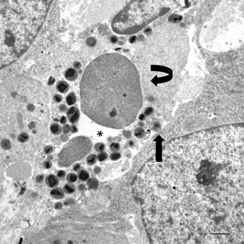
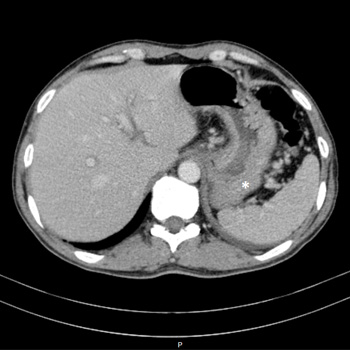
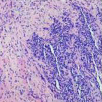
A case of poorly cohesive NOS gastric carcinoma, characterised by high-grade tumour-associated tissue eosinophilia (TATE), is studied by transmission electron microscopy. Eosinophil clustering around single tumour cells constituted a recurrent ultrastructural hallmark. Some eosinophils were in intimate contact with tumour cells and exhibited extracellular trap cell death (ETosis): a non-apoptotic cell death process, recently described in non-neoplastic, eosinophil-associated diseases. Discharge of chromatin material and specific granules, due to eosinophil ETosis, was polarised towards single tumour cells that showed various degrees of cytopathogenic changes. Our data suggest that eosinophil ETosis may exert an antitumoural activity in gastric cancer.
|
Views: 542
HTML: 70
PDF: 311
|
Overall gastric cancer incidence is decreasing, but incidence of gastric signet ring cell carcinoma has been rising. The diagnosis can be challenging. It has a poorer prognosis because it tends to be diagnosed at advanced stages. Lymphedema is a rare presentation. We report a rare presentation of signet ring cell carcinoma in a 49-year old male, with no underlying medical condition. The patient presented with lymphedema of lower limbs, scrotum and abdominal wall.
|
Views: 358
HTML: 48
PDF: 344
|
Central nervous system (CNS) lymphoma is a rare and aggressive primary neoplasm that comprises a small proportion of brain tumours and non-Hodgkin lymphomas. We present a case report of a 64-year-old woman with CNS lymphoma, who exhibited cognitive changes, weight loss and neurological symptoms. Imaging scans revealed multiple lesions in the brain and thrombosis in the venous sinuses. A diagnosis of diffuse large B-cell lymphoma of the CNS was confirmed through histological examination. The patient underwent treatment with corticosteroids and chemotherapy, but experienced clinical deterioration with thrombocytopenia and disease progression. Despite efforts to manage complications and provide targeted therapy, the patient passed away. Primary CNS lymphoma typically responds well to chemotherapy, and prognostic factors such as age and functional status play a significant role in patient outcomes. However, complications such as thromboembolism pose challenges during treatment due to the hypercoagulable state induced by chemotherapy agents. The pathophysiology of thromboembolic events in the context of malignancy remains uncertain but may involve direct tumour compression, vascular invasion and alterations in coagulation factors. The diagnostic process for CNS lymphoma can be complex, and the information obtained from cerebrospinal fluid analysis, including flow cytometry, may be limited in cases with low cell counts. Ongoing research exploring genetic tests and biomarkers shows promise for improving diagnostic accuracy in such cases. This case underscores the need for comprehensive management strategies that address both the neoplasm and its associated complications, to optimise patient outcomes.
|
Views: 296
HTML: 104
PDF: 361
|
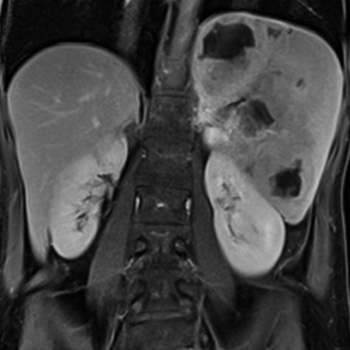
Primary splenic lymphoma (PSL) is a rare disease and an improbable cause of splenomegaly or splenic nodules. On the contrary, splenic secondary involvement as part of an advanced lymphoproliferative disorder is more common.
The authors present the case of a 49-year-old woman with a primary splenic diffuse large B-cell lymphoma (PS-DLBCL), in which the absence of other organs’ involvement determined an ultrasound-guided biopsy of the spleen to achieve a definitive diagnosis.
With this case the authors intend to emphasise the extensive differential diagnosis of splenomegaly, splenic nodules or infiltrates, the usefulness of splenic biopsy in establishing the diagnosis and recall a rare disease, with non-specific presenting symptoms, in which the diagnostic workup is challenging.
|
Views: 369
HTML: 45
PDF: 418
|
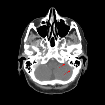
Introduction: Rhabdomyosarcoma is a high-grade malignant neoplasm with skeletal muscle differentiation; a common soft tissue sarcoma in children but considered one of the rarest in adults.
Case description: We report a case of 35-year-old male with a chronic productive cough and haemoptysis for five days. A CT scan of the nasopharynx revealed a blocked left maxillary and ethmoid sinus with bone destruction. These findings raised a suspicion of a tumour, and trans-nasal endoscopic sinus surgery was performed.
Discussion: Nasal rhabdomyosarcoma is a rare adult malignant tumour. Most patients have lymphatic metastasis or skull base tumour infiltration at the time of the initial diagnosis and treatment, which poses a challenge to the diagnosis and management.
Conclusion: Nasal acinar rhabdomyosarcoma, one of the histopathological types of rhabdomyosarcoma, has rapid disease progression and high mortality. Therefore, the treatment of rhabdomyosarcoma requires a combination of surgery, chemotherapy, radiotherapy, and immunotherapy to work together to achieve the best care for the patient.
| 2.1 = | 1.762 Cit. to date |
| 842 Docs. to date |


Publisher
Official Journal of the
European Federation of Internal Medicine
www.efim.org
Publisher: SMC media Srl
Via Giovenale, 7 - 20136 Milan - Italy
P.IVA 07626490960
info@ejcrim.com
www.ejcrim.com - ISSN: 2284-2594 - © EFIM 2014-2024, Published by SMC Media srl, Italy - Privacy policy

