EJCRIM 2023 CiteScore
| 2.1 = | 1.762 Cit. to date |
| 842 Docs. to date |
Last updated on 05 May, 2024
Updated monthly
Updated monthly
Powered by


|
Views: 129
HTML: 10
PDF: 127
|
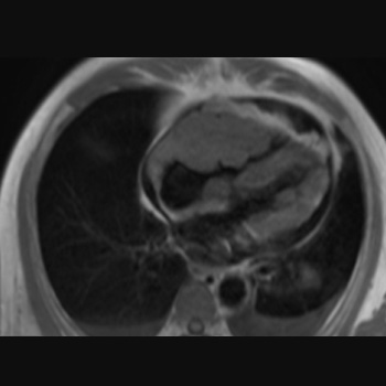
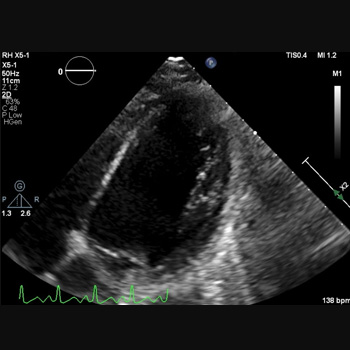
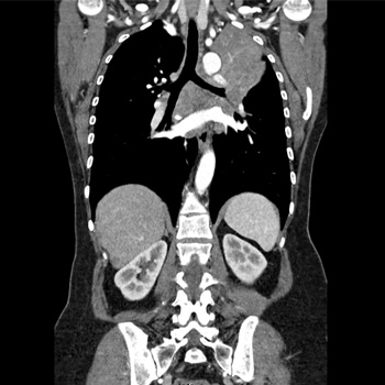
Background: This study presents a patient diagnosed with tricuspid valvular stenosis due to right ventricular lymphoma, who was treated successfully.
Case presentation: A 66-year-old man with a history of worsening shortness of breath during activity for the last three weeks sought medical attention. The patient later experienced swelling in the extremities, fluid build-up around the lungs and abdominal fluid accumulation, with no reported chest pain, fever, or weight loss. An echocardiogram found a mass in the lateral wall near the tricuspid valve of the right ventricle, leading to moderate tricuspid stenosis. The cardiac magnetic resonance imaging (MRI) revealed a lumpy, poorly defined mass that invaded the heart muscle and displayed varied enhancement after contrast administration. Suspicion arose for a malignant tumour or metastatic lesion due to its features and contrast uptake capability. A percutaneous biopsy was carried out on the mass in the right ventricle to confirm the diagnosis. The pathology report indicated a diagnosis of non-Hodgkin’s lymphoma. After being diagnosed, the patient underwent chemotherapy using the R-CHOP regimen. Over time the symptoms improved, and echocardiograms revealed a decrease in the size of the tumour. After undergoing six rounds of chemotherapy, a cardiac MRI four months later showed no signs of a tumour. After that, the patient resumed their regular activities.
Conclusion: Right ventricular tumours are mostly malignant lesions and often have an inferior prognosis. Early diagnosis with imaging techniques and myocardial biopsy is necessary to deliver life-saving treatment quickly.
|
Views: 421
HTML: 37
PDF: 274
|
Introduction: Combination-based adjuvant chemotherapy utilising capecitabine and oxaliplatin is widely used in gastric cancer treatment. Rare but severe cardiac events such as prolonged QT, cardiac arrest and cardiogenic shock can result from their use.
Case description: A 45-year-old female with gastric adenocarcinoma was started on capecitabine-oxaliplatin chemotherapy one week before presenting to the emergency department with weakness. Blood pressure was 78/56 mmHg, heart rate 140 bpm and oxygen saturation 85%. She became unresponsive with pulseless ventricular fibrillation; CPR was initiated with immediate intubation. She received two shocks with a return of spontaneous circulation. Laboratory tests revealed serum potassium (3.1 mmol/l), magnesium (1.1 mg/dl) and troponin (0.46 ng/ml). An EKG revealed sinus tachycardia with a prolonged QT interval (556 ms). The combined effects of capecitabine, oxaliplatin and electrolyte abnormalities likely contributed to the QT prolongation. An echocardiogram demonstrated an ejection fraction of 10%–15%. An emergent right-heart catheterisation showed right atrial pressure of 10 mmHg and pulmonary artery pressure of 30/18 mmHg; cardiac output and index were not recorded. An intra-aortic balloon pump was placed, and she was admitted to the ICU for cardiogenic shock requiring norepinephrine, vasopressin and dobutamine. A repeat echocardiogram showed a significantly improved ejection fraction of 65%, and she was discharged.
Discussion: Capecitabine and oxaliplatin cardiotoxicity is an exceedingly rare occurrence, with both drugs reported to cause QT prolongation.
Conclusion: Healthcare providers must recognise the QT prolongation effects of capecitabine and oxaliplatin, leading to life-threatening cardiac arrhythmias.
|
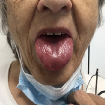
A case of amyloid goitre in heavy chain amyloidosis: diagnostic challenges and clinical implications
Views: 204
HTML: 28
PDF: 178
|
Immunoglobulin heavy chain amyloidosis (AH amyloidosis) is an extremely rare subtype of immunoglobulin-derived amyloidosis and there is limited literature on how to diagnose and manage this disorder. We describe a rare case of AH amyloidosis with amyloid goitre and the importance of mass spectrometry in the identification of the different types of amyloids. While additional studies are needed, several observations suggest important practical implications, including differences in clinical picture, prognosis, and pathologic diagnosis.

|
Views: 294
HTML: 52
PDF: 201
|
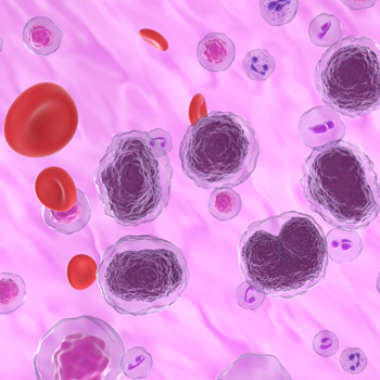
Chronic lymphocytic leukaemia (CLL) is a lymphoproliferative disorder characterised by an accumulation of monoclonal B lymphocytes, with an increased risk of secondary cancers. The coexistence of CLL and chronic myeloid leukaemia (CML) is a rare phenomenon, with three main types being classified: CML preceding CLL, CLL preceding CML and simultaneous occurrence. The coexistence of these chronic leukaemias poses a complex clinical challenge, with the underlying mechanisms of their association remaining enigmatic. Here, we present a report of an elderly male with a long history of CLL, who was subsequently diagnosed with secondary CML.
|
Views: 610
HTML: 18
PDF: 365
|
Introduction: During treatment for malignant lymphoma, cytopenia can develop for several reasons. This can range from mild cytopenias leading to infection and bleeding to full-blown drug-induced aplastic anaemia. While aplastic anaemia affects individuals of all genders and ages, here, we describe aplastic anaemia after chemotherapy exposure to bendamustine in a 65-year-old female with non-Hodgkin’s lymphoma.
Case description: A 65-year-old woman with recurrent indolent marginal zone lymphoma and post-chemotherapy with bendamustine and rituximab, presented with a neutropenic fever and was admitted with a leading diagnosis of sepsis. In the previous two weeks, the patient required regular transfusions of packed red blood cells and platelets and maintained a daily ZARXIO® regimen. Laboratory results revealed pancytopenia, and broad-spectrum antibiotics (cefepime/vancomycin) were given. The patient was subsequently admitted to the hospital under the care of the haematology/oncology team and was ultimately diagnosed with aplastic anaemia, likely as a consequence of bendamustine chemoimmunotherapy. She elicited a positive response to the triple immunosuppressive therapy (IST) regimen (two immunotherapeutic agents plus one anti-thymocyte globulin (ATG), after which her cell counts returned to normal.
Conclusions: This case underscores the importance of recognising haematologic complications linked to bendamustine and advocates for further research to increase the understanding among healthcare professionals of drug-induced aplastic anaemia. Bendamustine can cause severe autoimmune haemolytic anaemia and aplastic anaemia and may require multiple transfusions and a multidrug regimen for treatment. The use of ATG as a therapeutic intervention is appropriate because it has been effective in treating aplastic anaemia.
|
Views: 453
HTML: 65
PDF: 370
|
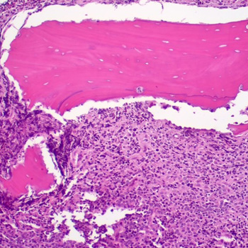
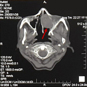
Background: Primary bone lymphoma (PBL) is a rare form of extra nodal non-Hodgkin’s lymphoma (NHL).
Case description: We describe a 39-year-old-male with no medical history who presented with unilateral facial swelling following a tooth extraction. Initial diagnoses after various presentations over the course of three weeks, based on inflammatory and infectious aetiologies. However, the patient was ultimately diagnosed with diffuse large B-cell lymphoma.
Discussion: Symptoms of PBL are very similar to inflammatory and infectious diseases of the bone, such as osteomyelitis or osteonecrosis. Clinical features of PBL involving the head and neck include persistent jaw pain, tooth mobility secondary to extensive destruction of bone, and in advanced cases, lip numbness and swelling. On examination it may present as an exposed necrotic bone with surrounding soft tissue oedema. Misdiagnosis of these lesions as an infectious or inflammatory aetiology may lead to an unnecessary delay in lymphoma treatment, and subsequently worsen the prognosis if caught at a later stage. Therefore, any concerning lesion, especially in the oral cavity, must be subjected to early histopathological evaluation to differentiate PBL from osteomyelitis and/or osteonecrosis.
Conclusion: This case report highlights the importance of an early histopathological evaluation to prevent delay in the diagnosis of primary bone lymphomas.
|
Views: 553
HTML: 54
PDF: 334
|
A nuclear protein in testis (NUT) midline carcinoma arises from squamous cells and is often located in the head, neck, and lungs. This report focuses on the negative p63 mutation and older age at the diagnosis of a NUT carcinoma, which has significant prognostic implications. A 62-year-old patient presented initially with a three-year history of recurring frontal headaches, intermittent nasal bleeding, and a sensation of a nasal cavity mass. An incisional biopsy revealed a poorly differentiated NUT carcinoma in the left maxillary sinus. A functional endoscopic sinus surgery was performed, but the cancer recurred. As a result, a total maxillectomy was performed, and the patient was declared cancer-free with no evidence of residual disease. This is a rare instance of a p63-negative midline NUT cell carcinoma (NCC) in an elderly patient, which could potentially contribute to a more favourable prognosis and longer survival compared to other reported cases.
|
Views: 594
HTML: 23
PDF: 374
|
Introduction: Most lung cancers are diagnosed at an advanced stage. Common metastatic sites include the brain, bone, liver and adrenal glands. Ocular metastases, however, are extremely rare. We present a case of advanced lung adenocarcinoma presenting exclusively with photopsias attributable to retinal metastases.
Case description: We describe a woman in her fifties, a lifetime non-smoker with an unremarkable medical and family history, who presented to the emergency department with photopsias for a week. Ophthalmology evaluation revealed decreased visual acuity bilaterally, and a fundus examination disclosed lesions suggestive of bilateral retinal metastases. A comprehensive evaluation diagnosed a stage IVb lung adenocarcinoma with exon 19 mutation on epidermal growth factor receptor gene. Subsequently, she developed complaints of headaches and dizziness. She received frontline osimertinib 80 mg daily, preceded by upfront whole-brain radiation therapy with partial orbital inclusion for symptomatic ocular and brain metastases. After ten radiation therapy sessions, her complaints were resolved and an ophthalmology revaluation revealed improvement in visual acuity and resolution of photopsia complaints. The patient is currently on osimertinib and preserves an ECOG score of 0.
Conclusion: Retinal metastases usually indicate advanced disease, so presenting with isolated ocular symptoms is exceedingly rare. Especially in cases of uncommon metastases, a multidisciplinary approach is fundamental for a prompt diagnosis and timely treatment, impacting prognosis and quality of life.

|
Views: 373
HTML: 28
PDF: 292
|
Immunoglobulin G4-related disease (IgG4-RD) is an autoimmune disease characterized by elevated serum IgG4 levels. It has the potential to affect multiple organs. Despite the diverse manifestations of IgG4-RD, the association with coronary artery disease (CAD) remains poorly understood due to limited evidence. We report the case of a 52-year-old male patient who exhibited typical angina upon exertion, accompanied by elevated serum IgG4 levels. Coronary computed tomographic angiography (CCTA) revealed the presence of pseudotumor formations surrounding and aneurysm changes affecting all coronary arteries, consistent with IgG4-RD. The patient was treated with prednisolone and azathioprine, with the possibility of additional rituximab therapy if symptomatology failed to improve. This case sheds light on the rare occurrence of IgG4-RD with coronary artery involvement and underscores the importance of recognizing this unique clinical entity for appropriate management and further research.

|
Views: 668
HTML: 80
PDF: 536
|
Sodium-glucose cotransporter-2 (iSGLT2) inhibitors, which include dapagliflozin, canagliflozin and empagliflozin, are a class of drugs initially used in the oral treatment of diabetes, heart failure and renal failure. They target the reabsorption of glucose in the kidney. Although they bring benefit to patients with these conditions and in general produce few adverse effects, in some cases, iSGLT2 can cause serious adverse effects such as metabolic acidosis, and fungal or bacterial urinary infections. Oncology patients, who in general have a weak immune system and are usually treated with chemotherapy and/or immunotherapy, are more susceptible to this type of adverse events than other patients. For this reason, it is necessary to adequately select the patients eligible to receive this type of drug and evaluate the potential benefits for them. In this series of five cases, we present two cases of metabolic acidosis, two cases of bacterial urinary sepsis, and one case of fungal urinary sepsis that occurred in patients admitted to the Medical Oncology Department of the University Hospital of Salamanca in 2023.
| 2.1 = | 1.762 Cit. to date |
| 842 Docs. to date |


Publisher
Official Journal of the
European Federation of Internal Medicine
www.efim.org
Publisher: SMC media Srl
Via Giovenale, 7 - 20136 Milan - Italy
P.IVA 07626490960
info@ejcrim.com
www.ejcrim.com - ISSN: 2284-2594 - © EFIM 2014-2024, Published by SMC Media srl, Italy - Privacy policy

