EJCRIM 2023 CiteScore
| 2.1 = | 1.762 Cit. to date |
| 842 Docs. to date |
Last updated on 05 May, 2024
Updated monthly
Updated monthly
Powered by


|
Views: 0
HTML: 0
PDF: 0
|
Background: Acute pancreatitis is a common cause of hospitalisation characterised by inflammation of the pancreas. While mechanical, toxic and iatrogenic factors typically cause it, post-oesophagogastroduodenoscopy (EGD) pancreatitis is extremely rare. This report examines a case of acute pancreatitis following EGD, aiming to highlight this rare but significant complication.
Case description: A 46-year-old woman with a history of breast cancer, anxiety, vitamin D deficiency and gastro-oesophageal reflux disease underwent an EGD, which revealed and led to the removal of duodenal polyps. Six hours post-procedure, she presented with severe abdominal pain radiating to her back, accompanied by nausea. Laboratory results indicated elevated lipase levels, and a computed tomography (CT) scan confirmed acute pancreatitis. The patient was managed with aggressive fluid resuscitation, bowel rest and pain management, leading to an improvement in her condition and subsequent discharge. We believe that the pancreatitis was likely caused by the use of cautery during the endoscopic mucosal resection of duodenal polyps.
Conclusion: This case underscores the need for clinicians to recognise acute pancreatitis as a potential complication of EGD, especially in the absence of other common risk factors.

|
Views: 23
HTML: 0
PDF: 13
|
This case report details the complex diagnostic odyssey of a 60-year-old female grappling with chronic liver disease, initially diagnosed with hepatic encephalopathy (HE). Despite initial treatment with lactulose and rifaximin, her neurological symptoms worsened, leading to the identification of concurrent acquired hepatocerebral degeneration (AHD). This condition is characterised by cognitive decline, movement disorders and distinctive imaging abnormalities. The discussion highlights the challenges in distinguishing AHD from HE, underscoring the sophisticated diagnostic and management strategies required for such intricate cases in the realm of chronic liver disease.

|
Views: 38
HTML: 5
PDF: 29
|
Introduction: Primary squamous cell carcinoma of the liver (SCC) is a rare and challenging pathology. As an aggressive cancer, the prognosis is extremely poor with less than 12 months overall survival. In view of its low prevalence, we report the case of an elderly patient with primary squamous cell carcinoma of the liver.
Case description: A 74-year-old female, with no pathological history, presented with acute right hypochondrium pain associated with shivering, asthenia and weight loss. We diagnosed primary hepatic squamous cell carcinoma by pathological analysis.
Conclusion: Primary hepatic squamous cell carcinoma represents a rare malignant tumour with extremely poor prognosis. There is no established treatment protocol for this disease and a multidisciplinary approach is needed to choose the best therapeutic option.

|
Views: 119
HTML: 18
PDF: 62
|
Background: Primary hepatic epithelioid hemangioendothelioma (HEHE) is an extremely rare tumour of vascular origin with an incidence of <0.1 cases per 100,000 people worldwide.
Case description: A 29-year-old female with the history of epigastric pain and unintentional weight loss (3 kg over six months) was referred for upper endoscopy. The examination was without visual pathological findings, but a rapid urease test was positive. First-line treatment with clarithromycin-containing triple therapy for Helicobacter pylori infection was given. After completion of eradication therapy, diffuse abdominal pain developed. An abdominal computed tomography (CT) showed multiple liver nodules. Three consecutive core liver biopsies were performed and were inconclusive. A subsequent surgical liver nodule resection was performed. Histopathology of the specimen revealed grade 2 hepatocellular carcinoma; bone scintigraphy was negative for metastasis. A multidisciplinary team (MDT) recommended giving the patient sorafenib, which was poorly tolerated. The histology was reviewed using immunohistochemistry staining at the request of the oncologist, which showed expression of CD31 and CD34. Based on clinical, morphological and immunohistochemistry findings, a diagnosis of hepatic epithelioid hemangioendothelioma was made. Based on the multidisciplinary team's findings, liver transplantation was indicated as the only curative treatment.
Conclusion: Because of the rarity of this disease, combining clinical, radiological and histopathological methods as well as an MDT approach can help to reach the correct final diagnosis. As demonstrated in this clinical case, it is crucial to perform immunohistochemistry of a liver biopsy to confirm a HEHE diagnosis.

|
Views: 234
HTML: 10
PDF: 93
|
Hepatitis A is a mild self-limiting infection of the liver with spontaneous resolution of symptoms in most cases. However, clinicians should be aware of some commonly encountered complications and extrahepatic manifestations associated with hepatitis A for timely diagnosis and treatment. Rhabdomyolysis, an exceedingly rare complication of hepatitis A, is scarcely documented. We present a case of a 64-year-old man with symptoms consistent with rhabdomyolysis and an evanescent rash secondary to acute hepatitis A. He eventually recovered with conservative management. This case emphasizes the importance of recognizing and treating atypical presentations of acute hepatitis A infection.

|
Views: 154
HTML: 21
PDF: 81
|
Khat is a plant that is commonly used for its stimulating effects and is chewed for its psychoactive properties. It creates feelings of euphoria that are similar to when taking amphetamines. There is an association between khat and liver injury, but the mechanism is not well known. We present three cases of khat-induced liver injury. All cases have elevated IgG and either positive antinuclear antibodies (ANA) or anti-smooth muscle antibody (ASMA); each case has a different course and requires different management. One case improved only by stopping khat, one required a short course of steroids and the last case required treatment such as that for autoimmune hepatitis (AIH).

|
Views: 173
HTML: 12
PDF: 146
|
Late onset combined immunodeficiency (LOCID) is a rare variant of common variable immunodeficiency (CVID), typically affecting adult patients who present with opportunistic infections (OI) and/or low CD4+ T lymphocytes. Diagnostic delay is common due to the rareness of this entity, increasing morbidity and mortality. We report on a 66-year-old male who developed a severe gastrointestinal cytomegalovirus (CMV) infection, refractory to antiviral treatment and anti-cytomegalovirus specific human immunoglobulin administration, with a fatal outcome due to an undiagnosed LOCID.

|
A rare case of biloma after ascending cholangitis and endoscopic retrograde cholangiopancreatography
Views: 238
HTML: 29
PDF: 139
|
Introduction: Biloma is an uncommon form of liver abscess composed of bile usually associated with procedures of the biliary tree and gallbladder. Cholangitis can be acute or chronic, can result in partial or complete obstruction of the flow of bile. The infection of the bile is so common, that positive blood cultures are highly characteristic. In the case of a suppurative cholangitis with signs of sepsis treatment alone with antibiotics is usually not sufficient to achieve medical remission. Multiple hepatic abscesses are often present, and the mortality approaches 100% unless prompt endoscopic or surgical relief of the obstruction and drainage of infected bile are carried out. Endoscopic retrograde cholangiopancreatography ERCP with endoscopic sphincterotomy is the preferred initial procedure for both establishing a definitive diagnosis and providing effective therapy.
Case description: We present the case of a 69-year-old female patient with complex chronic comorbidities who presented with acute cholangitis initially managed with endoscopically inserted stent and later complicated by sepsis and biloma formation. The bile was drained, and it showed an infection with Candida spp. requiring antifungal therapy.
Conclusions: The failure to perform sphincterotomy in patients with suppurative cholangitis can contribute to the backflow of bile and worse outcomes.
|
Views: 195
HTML: 12
PDF: 152
|
Introduction: Caustic substances ingestion results in a complex syndrome. The patient characteristics and severity of injury are important prognostic predictors. The monitoring of clinical changes and the multidisciplinary approach are necessary to prevent death in the early stages of the poisoning.
Case description: The case report describes the suicide of a woman by ingestion of a large amount of 15% sulfuric acid for suicidal purposes (15–20 ml). The initial conditions were stable, and no changes were found on a CT scan. However, the main sign was a severe metabolic acidosis. After 7 hours, haematemesis and oedema of the larynx appeared, and oro-tracheal intubation and ICU admission were necessary. Consequent progressive haemodynamic deterioration with persistent severe metabolic acidosis, increasing lactates and septic shock appeared. A new CT scan with contrast was performed 22 hours later detecting diffuse perforations and liquid in pleurae and abdomen. A pleural sample showed necrotic liquid. The death was 24 hours after ingestion and no surgical treatment was performed because of the diffuse lesions to the thoracoabdominal districts.
Conclusions: Early detection of gastroenteric lesions and the monitoring of clinical changes are mandatory to avoid the death of the patient. Gastroenteric perforations require an immediate radiological evaluation and surgical treatment. The clinical case report states the severity of prognosis was related to high doses of sulfuric acid ingestion. The immediate metabolic acidosis is related to quick subsequent severe systemic pathological lesions of the gastrointestinal tract. The severity of absorption metabolic acidosis, consequently, may be an early and prognostic sign of the late chest and abdominal lesions.
|
Views: 226
HTML: 35
PDF: 169
|
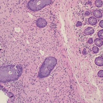
Schwann cells are found in the peripheral nervous system and can sometimes appear as benign hamartoma lesions in various parts of the body. Although rare in the gastrointestinal (GI) tract, they have been observed in the colon.
Recently, mucosal Schwann cell hamartomas of the GI tract have been studied, and it was discovered that they had yet to be investigated up to 2009. In this context, we present the case of a 60-year-old man who was found to have lesions in the transverse colon during a routine colonoscopy. No further investigations were conducted since these lesions have not been associated with any risk of malignancy transformation and have not been linked to any inherited syndromes.
| 2.1 = | 1.762 Cit. to date |
| 842 Docs. to date |


Publisher
Official Journal of the
European Federation of Internal Medicine
www.efim.org
Publisher: SMC media Srl
Via Giovenale, 7 - 20136 Milan - Italy
P.IVA 07626490960
info@ejcrim.com
www.ejcrim.com - ISSN: 2284-2594 - © EFIM 2014-2024, Published by SMC Media srl, Italy - Privacy policy

