EJCRIM 2023 CiteScore
| 2.1 = | 1.762 Cit. to date |
| 842 Docs. to date |
Last updated on 05 May, 2024
Updated monthly
Updated monthly
Powered by


|
Views: 211
HTML: 105
PDF: 202
|
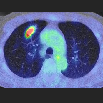
A 69-year-old man was diagnosed with lung adenocarcinoma with metastasis because two masses in the right intercostal space and right back muscle showed high accumulation on positron emission tomography (PET). The 6-month treatment with osimertinib significantly reduced his lung lesion, but no changes were observed in the metastatic lesions. Needle biopsy revealed that the lesion in the right back muscle was a schwannoma. Surgical resection revealed that the right intercostal lesion was also a schwannoma; subsequently, a right upper lobectomy was performed. The patient was finally diagnosed with lung adenocarcinoma without metastasis. High accumulations of lesions observed on PET may indicate schwannomas.

|
Views: 264
HTML: 33
PDF: 260
|
|
Views: 394
HTML: 67
PDF: 341
|
Tracheoesophageal prosthesis (TEP) is an artificial connection between the trachea and esophagus allowing air into the upper esophagus from the trachea thereby vibrating it. TEPs give patients who lose their vocal cords to laryngectomies a tracheoesophageal voice. A potential complication of this is silent aspiration of gastric content. We present a case of a 69-year-old female with a TEP placed after a laryngectomy for laryngeal cancer who presented to the hospital with shortness of breath and hypoxia. She was initially treated for a presumed diagnosis of chronic obstructive pulmonary disease (COPD) and congestive heart failure (CHF) exacerbations but continued to be hypoxic despite aggressive medical management. Further evaluation revealed silent aspirations as a consequence of TEP malfunction. Through our case report we urge clinicians to consider this differential diagnosis, as the clinical presentation of silent aspiration among patients with a TEP can be easily mistaken for a COPD exacerbation. A large number of patients with TEPs are smokers with underlying COPD.
|
Views: 367
HTML: 820
PDF: 375
|
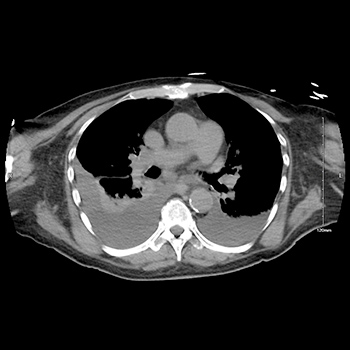
Chest pain and dyspnoea are among the most common complaints seen in the emergency room and each symptom calls for a broad differential diagnosis. Large hiatal hernias are infrequent, but they can lead to atypical symptoms mimicking different cardiovascular, pulmonary and neoplastic diseases. We present two cases of older patients with an apparent left atrial mass on transthoracic echocardiography, which was subsequently identified as hiatal hernia by other imaging modalities. A multidisciplinary team with multimodality imaging is necessary for diagnostic work-up of chest pain and dyspnoea of non-cardiac origin and especially for a suspected mass compressing the heart, causing chest discomfort.
|
Views: 355
HTML: 45
PDF: 357
|
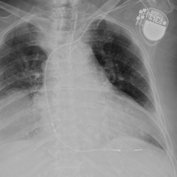
Intrathoracic kidney is a very rare finding, representing less than 5% of all renal ectopias. Because of the location of the liver, thoracic kidney on the right side is much less common than thoracic kidney on the left side. Although an increasing number of case reports are being published in the literature, few describe the impact of the ectopia on kidney function. We report the case of a woman with intrathoracic right kidney and chronic kidney disease that was initially misdiagnosed as pneumonia because of its presentation on chest x-ray. We highlight the need to including this condition in the differential diagnosis as the literature rarely links it to changes in kidney function.
|
Views: 419
HTML: 98
PDF: 340
|
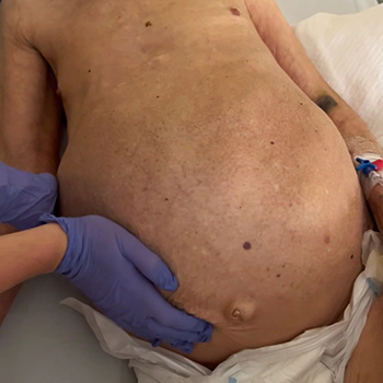
Caroli disease is a rare congenital pathology caused by mutation of the PKHD1 gene (polycystic kidney and hepatic disease 1), also responsible for autosomal recessive polycystic kidney disease. Characterized by segmental and multifocal dilatation of the large intrahepatic bile ducts, classic disease involves only malformation of the biliary tract. The association with congenital hepatic fibrosis is called Caroli syndrome. We describe the case of an 84-year-old man with Caroli syndrome diagnosed in 1997 by liver biopsy. The CT scan revealed massive hepatomegaly, extending to the pelvic region, and almost total replacement of the parenchyma by numerous cystic formations, no evidence of bile duct dilatation, and no ascites or splenomegaly suggestive of portal hypertension. The atypical clinical presentation, with no reported complications, resembles that of a space-occupying lesion with an indolent course, previously misdiagnosed as metastatic neoplasm.
|
Views: 490
HTML: 199
PDF: 444
|
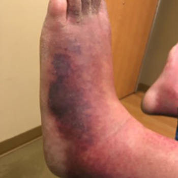
Livedoid vasculopathy (LV) is a rare clinical condition presenting as painful lesions mostly on the lower extremities. We present a case of LV with peripheral neuropathy in a young man initially misdiagnosed and treated for cellulitis. He was started on aspirin, pentoxifylline and apixaban immediately after the diagnosis of LV. However, pain management was a real challenge for the clinicians. Hence, he was later treated with epoprostenol and amlodipine for vasodilation, steroids for any possible inflammation, and antibiotics to treat superimposed infection. Irrespective of all the above, his pain was uncontrollable, and he finally received ketamine infusions along with narcotics, achieving better pain control. Various studies support the use of intravenous immunoglobulin and anti-TNF agents for pain relief in idiopathic and secondary LV. Intermittent low-dose dabigatran has also been found to be effective in the maintenance of remission in LV. However, no large studies have yet been conducted to confirm the efficacy of these medications.
| 2.1 = | 1.762 Cit. to date |
| 842 Docs. to date |


Publisher
Official Journal of the
European Federation of Internal Medicine
www.efim.org
Publisher: SMC media Srl
Via Giovenale, 7 - 20136 Milan - Italy
P.IVA 07626490960
info@ejcrim.com
www.ejcrim.com - ISSN: 2284-2594 - © EFIM 2014-2024, Published by SMC Media srl, Italy - Privacy policy

