EJCRIM 2023 CiteScore
| 2.1 = | 1.762 Cit. to date |
| 842 Docs. to date |
Last updated on 05 May, 2024
Updated monthly
Updated monthly
Powered by


|
Views: 404
HTML: 53
PDF: 269
|
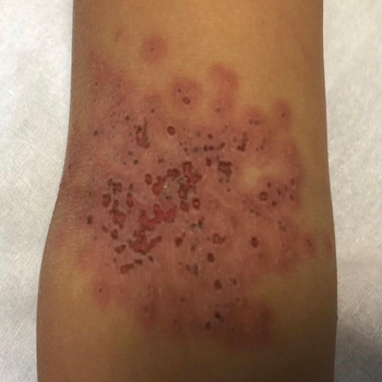
Introduction: Kaposi’s varicelliform eruption (KVE), also known as eczema herpeticum or eczema vaccinatum, is an acute dermatosis that affects patients with chronic dermatopathies. The diagnosis is primarily clinical and is characterised by the presence of a vesicular exanthema on physical examination. The exanthema subsequently evolves into crusted lesions with typical circular ulcerations in ‘punched-out’ areas on the skin affected by the underlying dermatopathy.
Case description: We present the case of a 6-year-old patient who presented to the Paediatric Emergency department with skin lesions consistent with eczema herpeticum. The patient’s management was initially outpatient; however, due to the slow progression of the condition, hospitalisation and intravenous antiviral treatment were initiated.
Discussion: KVE affects patients with chronic dermatoses, especially atopic dermatitis. It is important to know the clinical presentation for an early suspicion. KVE is a medical emergency that requires prompt diagnosis and treatment. It can progress to secondary viraemia, which can be fatal in up to 10% of immunocompetent individuals and up to 50% of immunocompromised individuals. It is important to be aware of this condition and to start early treatment with antivirals, especially given the high prevalence of atopic dermatitis in our population. This condition is one of the most serious complications that can occur in these patients.

|
Views: 96
HTML: 20
PDF: 110
|
Introduction: Rectus sheath haematoma (RSH) has become increasingly common but is often underdiagnosed. Prompt diagnosis will avoid unnecessary investigations and procedures, resulting in early treatment and a better outcome.
Case description: We described a case of a spontaneous RSH with intraperitoneal extension and formation of a vesico-haematoma fistula, which was initially misdiagnosed as a urinary tract infection. The diagnosis was made ten days after admission, when a CT scan showed an over-16 cm RSH with intraperitoneal extension, bladder perforation and a vesico-haematoma fistula. The patient was managed conservatively.
Discussion: RSH accounts for less than 2% of acute abdomen cases and is often unrecognised. Its presentation can mimic other intra-abdominal pathologies, and the diagnosis is often delayed or missed. Complications can arise from an RSH although it is generally viewed as a self-limiting condition.
Conclusion: RSH has become increasingly common, and we would like to highlight the need to include abdominal wall pathologies in the initial differential diagnoses of acute abdomen to avoid delay in diagnosis.
|
Views: 199
HTML: 21
PDF: 150
|
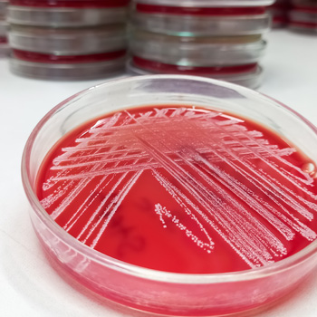
Kocuria kristinae is a Gram-positive commensal bacterium, rarely responsible for infection in immunocompromised patients.
A 29-year-old woman affected by intestinal pseudo-obstruction and requiring home parenteral nutrition, was hospitalised for fever and shivering during the infusion through a long-term central venous catheter (CVC).
Blood cultures were positive for K. kristinae infection. At a chest CT scan, two partially cavitated nodular lesions were evidenced. Meropenem antibiotic therapy was used locally and systemically, resulting in catheter use restoration.
A chest CT scan two months later at follow-up showed two centimetric, fibrotic and disventilatory areas replacing the previous nodular thickenings.
Kokuria kristinae was responsible for haematogenous pulmonary involvement with excavated nodules, requiring a differential diagnosis. Moreover, in the case of a CVC infection, in addition to the risk of right endocarditis, haematogenous pneumonia must also be considered.
|
Views: 245
HTML: 17
PDF: 236
|
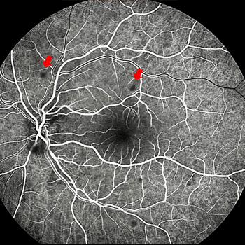
Introduction: A case of ocular bartonellosis under anti-tumour necrosis factor treatment is described.
Case description: A 29-year-old woman with psoriasis who had been on certolizumab treatment was examined with a left visual deterioration following a fever bout, malaise, and placoid erythematous rashes on her neck. As there was acute anterior uveitis in her left eye, it was recommended to stop certolizumab treatment for a possible infectious aetiology. However, her physician elected to continue the certolizumab treatment. Ten days later, the patient noticed further visual decline despite the topical steroid treatment. This time, there were scattered yellow-white small retinitis foci at the left posterior pole. Infectious agents were searched and while Bartonella henselae antibodies were negative for immunoglobulin M, the immunoglobulin G titre was 1/80. Clinical findings were improved with the systemic treatment of oral trimethoprim-sulfamethoxazole (160/800 mg twice daily for six weeks) and azithromycin (500 mg once daily for two weeks).
Discussion: Though extremely rare, ocular bartonellosis should be kept in mind in patients on anti-tumour necrosis factor treatment as rapid and accurate diagnosis may end up with an excellent visual outcome and full recovery.
|
Views: 265
HTML: 14
PDF: 217
|
Castleman’s disease (CD) and thrombotic thrombocytopenic purpura (TTP) are rare diseases that can affect the general population, especially those with HIV. Owing to their rarity, the association between CD and TTP remains insufficiently understood. In this study, we present a case of a 53-year-old patient with controlled HIV infection who presented with fever, lymphadenopathy, severe anaemia, and thrombocytopenia. After a series of tests, the diagnosis was concurrent human herpesvirus 8 (HHV8)-related multicentric CD (MCD) and TTP. Only four male patients were previously reported having this association, with HHV8 present in four and HIV in three patients, suggesting that coinfection with HHV8 and HIV is a pivotal factor in MCD with TTP occurrence.
|
Views: 247
HTML: 80
PDF: 295
|
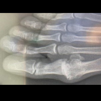
Background: In rare dermatology cases the differential diagnosis is challenging, e.g. when one nail is growing below another, the provisional diagnosis could be confusing. It may present as chronic paronychia, candidiasis, bacterial infections, rheumatoid arthritis, psoriasis, subungual tumours, or cysts.
Case description: We present a case of iatrogenic rupture of the nails of both big toes following a commonly known recommendation from physiotherapists in the initial stages of hallux valgus or chronic arthritis by using kinesio tape to prevent the big toe from fixation in the valgus position. The initial provisional diagnosis of retronychia was revised, and a final diagnosis of onychomadesis was made. The patient’s complaint was solved after around one year without any specific therapy.
Conclusion: The differential diagnosis for onychomadesis needs a careful and detailed history that may prevent a patient from a frightening diagnosis and painful, long-lasting treatments.
|
Views: 494
HTML: 42
PDF: 308
|
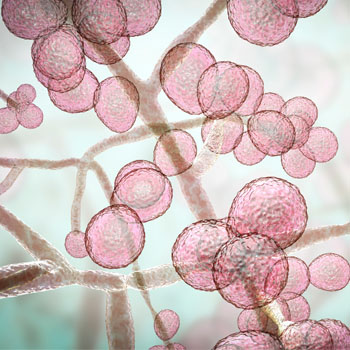
Infection caused by Candida auris has rapidly become a global health threat. C. auris created a significant healthcare burden due to various complicating factors, including misidentification by commercial identification methods, potent antifungal resistance, high mortality rates and the possibility of nosocomial outbreaks through direct contact. In Vietnam, there are currently no clinical reports on C. auris infections. Here, we present four clinical cases of C. auris infections in the Department of Pulmonary Medicine of Cho Ray Hospital in southern Vietnam. Through this report, we aim to highlight the attention to the emergence of C. auris in Vietnam. Further research on C. auris infections is warranted, focusing on newly observed clinical characteristics present in all cases in this report, including hypoalbuminaemia and corticosteroid usage. Moreover, one case of resistance to amphotericin B has been identified, possibly due to prior exposure to this antifungal agent.
|
Views: 254
HTML: 49
PDF: 214
|
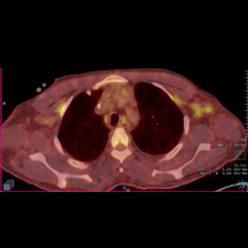
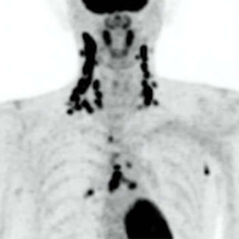
Kikuchi-Fujimoto disease (KFD), also called histiocytic necrotizing lymphadenitis, is more common in young women and typically presents with small, painful, localized cervical lymphadenopathy that resolves spontaneously within a few weeks. Laboratory findings are variable. As many as 40% of KFD cases are reported to be painless, and up to 22% to be generalized lymphadenopathy. Therefore, malignant lymphoma could be a differential diagnosis of KFD. A histopathologic diagnosis is needed when it is difficult to distinguish KFD from lymphoma. KFD typically shows small, highly accumulated cervical lymph nodes on fluorodeoxyglucose positron emission tomography (FDG-PET). This contrasts with malignant lymphoma, which tends to be associated with massive lymphadenopathy. In our case, a 40-year-old Japanese male presented with painless lumps in the right neck, accompanied by fever, night sweats, and loss of appetite. His symptoms and laboratory results worsened over a month. FDG-PET revealed highly accumulated uptake in cervical, mediastinal, and axillary lymph nodes. The PET imaging showed a small, high FDG uptake and contributed to the correct diagnosis of KFD. This case report highlights the importance of FDG-PET, which is a valuable diagnostic tool for KFD as it typically differentiates large clusters of small lymph nodes typical of KFD from normal lymph nodes.
|
Views: 443
HTML: 49
PDF: 260
|
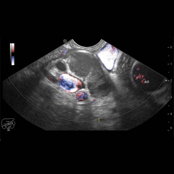
Tuberculosis (TB) is primarily a respiratory infection with huge mortality and morbidity worldwide. Extrapulmonary TB infection is common, affecting lymph nodes, pleura, and abdomen, but the primary biliary presentation without lung involvement is exceedingly rare. We report on a 38-year-old male patient who presented with isolated obstructive jaundice secondary to TB infection. This case highlights the importance of considering TB infection in the differential diagnosis of obstructive jaundice, especially in the endemic area. We also provide a literature review on TB infection, mainly in the biliary tract.
|
Views: 318
PDF: 241
HTML: 29
|
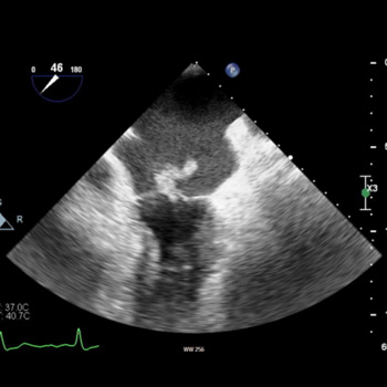
Introduction: There are very few documented cases of Escherichia coli endocarditis with cardiac abscesses in the literature. Here we describe a case presentation with diagnostic challenges and a multidisciplinary approach to management.
Case description: This is a rare presentation of E. coli endocarditis in a patient with a prosthetic aortic valve. Initial tests were inconclusive and further investigation with transoesophageal echocardiography was required to make the diagnosis. Despite initial improvement, the patient deteriorated and ultimately died of complications related to the presentation.
Discussion/conclusion: E. coli is a rare causative organism for endocarditis, which can itself be difficult to diagnose. A multidisciplinary approach to investigation and treatment is required when infective endocarditis is suspected. Transoesophageal echocardiography may be required to diagnose endocarditis when there is a strong clinical suspicion and risk factors present.
| 2.1 = | 1.762 Cit. to date |
| 842 Docs. to date |


Publisher
Official Journal of the
European Federation of Internal Medicine
www.efim.org
Publisher: SMC media Srl
Via Giovenale, 7 - 20136 Milan - Italy
P.IVA 07626490960
info@ejcrim.com
www.ejcrim.com - ISSN: 2284-2594 - © EFIM 2014-2024, Published by SMC Media srl, Italy - Privacy policy

