PET scanning may not distinguish benign schwannoma from metastasis in a patient with lung adenocarcinoma
  • Motoi Ugajin
    Department of Respiratory Medicine, Nagoya Tokushukai General Hospital, Kasugai City, Japan; Department of Respiratory Medicine and Allergology, Aichi Medical University, Nagakute City, Japan
  • Masakatsu Yamashita
    Department of Thoracic Surgery, Nagoya Tokushukai General Hospital, Kasugai City, Japan
  • Hisanori Kani
    Department of Thoracic Surgery, Nagoya Tokushukai General Hospital, Kasugai City, Japan

Keywords

Lung adenocarcinoma, schwannoma, positron emission tomography, biopsy

Abstract

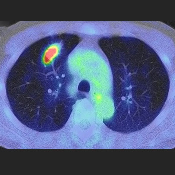
A 69-year-old man was diagnosed with lung adenocarcinoma with metastasis because two masses in the right intercostal space and right back muscle showed high accumulation on positron emission tomography (PET). The 6-month treatment with osimertinib significantly reduced his lung lesion, but no changes were observed in the metastatic lesions. Needle biopsy revealed that the lesion in the right back muscle was a schwannoma. Surgical resection revealed that the right intercostal lesion was also a schwannoma; subsequently, a right upper lobectomy was performed. The patient was finally diagnosed with lung adenocarcinoma without metastasis. High accumulations of lesions observed on PET may indicate schwannomas.

VIEW THE ENTIRE ARTICLE

References

  • Li J, Xu W, Kong F, Sun X, Zuo X. Meta-analysis: accuracy of 18FDG PET-CT for distant metastasis staging in lung cancer patients. Surg Oncol 2013;22:151–155.
  • Rami-Porta R, Call S, Dooms C, Obiols C, Sánchez M, Travis WD, et al. Lung cancer staging: a concise update. Eur Respir J 2018;51:1800190.
  • Ferner RE, O’Doherty MJ. Neurofibroma and schwannoma. Curr Opin Neurol 2002;15:679–684.
  • Dewey BJ, Howe BM, Spinner RJ, Johnson GB, Nathan MA, Wenger DE, et al. FDG PET/CT and MRI features of pathologically proven schwannomas. Clin Nucl Med 2021;46:289–296.
  • Kang S. Benign schwannoma mimicking metastatic lesion on F-18 FDG PET/CT in differentiated thyroid cancer. Nucl Med Mol Imaging 2013;47:138–140.
  • Boré P, Descourt R, Ollivier L, Le Roux P-Y, Abgral R. False positive 18F-FDG positron emission tomography findings in schwannoma – a caution for reporting physicians. Front Med (Lausanne) 2018;5:275.
  • Fujiuchi N, Saeki T, Takeuchi H, Sano H, Takahashi T, Matsuura K, et al. A false positive for metastatic lymph nodes in the axillary region of a breast cancer patient following mastectomy. Breast Cancer 2011;18:141–144.
  • Crist J, Hodge JR, Frick M, Leung FP, Hsu E, Gi MT, et al. Magnetic resonance imaging appearance of schwannomas from head to toe: a pictorial review. J Clin Imaging Sci 2017;7:38.
  • Hochhegger B, Marchiori E, Sedlaczek O, Irion K, Heussel CP, Ley S, et al. MRI in lung cancer: a pictorial essay. Br J Radiol 2011;84:661–668.
  • Hu S, Chen Y, Wang Y, Chen KM, Song Q. Clinical and CT manifestation of pleural schwannoma. Acta Radiol 2012;53:1137–1141.
  • Wang W, Cao K, Han Y, Zhu X, Ding J, Peng W. Computed tomographic characteristics of gastric schwannoma. J Int Med Res 2019;47:1975–1986.
  • Hu J, Liu X, Ge N, Wang S, Guo J, Wang G, et al. Role of endoscopic ultrasound and endoscopic resection for the treatment of gastric schwannoma. Medicine (Baltimore) 2017;96:e7175.
  • Li Q, Gao C, Juzi JT, Hao X. Analysis of 82 cases of retroperitoneal schwannoma. ANZ J Surg 2007;77:237–240.
  • Views: 211
    HTML downloads: 105
    PDF downloads: 202


    Published: 2023-08-31
    Issue: 2023: Vol 10 No 10 (view)


    How to cite:
    1.
    Ugajin M, Yamashita M, Kani H. PET scanning may not distinguish benign schwannoma from metastasis in a patient with lung adenocarcinoma. EJCRIM 2023;10 doi:10.12890/2023_004048.