Non-Hodgkin Lymphoma Causing Hypopituitarism Can Imaging Help Diagnosis and Management?
  • Uzma Khan
    Department of Endocrinology & Diabetes, University Hospitals Coventry & Warwickshire NHS Trust, UK https://orcid.org/0000-0002-5559-9300
  • Anton Borg
    Department of Haematology, South Warwickshire NHS Foundation Trust, UK
  • Radu Beltechi
    Department of Neurosurgery, University Hospitals Coventry & Warwickshire NHS Trust, UK
  • Hiten Mehta
    Department of Clinical Radiology, University Hospitals Coventry & Warwickshire NHS Trust, UK
  • Timothy Robbins
    Department of Endocrinology & Diabetes, University Hospitals Coventry & Warwickshire NHS Trust, UK
  • Harpal Randeva
    Department of Endocrinology & Diabetes, University Hospitals Coventry & Warwickshire NHS Trust, UK
  • Pratibha Machenahalli
    Department of Endocrinology & Diabetes, University Hospitals Coventry & Warwickshire NHS Trust, UK

Keywords

Non-Hodgkin lymphoma, CNS lymphoma, pituitary macroadenoma, sellar mass

Abstract

Non-Hodgkin lymphomas of the hypothalamus and pituitary are rare. They usually remain clinically silent until onset of compressive features affecting surrounding structures. When symptomatic, patients most commonly present with diabetes insipidus, headaches, ophthalmoplegia and/or bilateral hemianopia. 
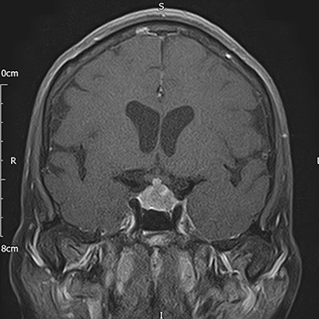
We report a case of a 67-year-old Caucasian female with a history of B-cell lymphoma in complete remission. She presented with left oculomotor nerve palsy and was subsequently found to have a sellar/suprasellar mass lesion on MRI. Alongside hypocortisolism and hypogonadotropic hypogonadism, she developed transient diabetes insipidus during her illness. Her clinical course was characterized by rapid intracranial progression of the sellar mass. MR spectroscopy suggested a diagnosis of lymphoma. Diagnostic biopsy confirmed high-grade diffuse large B-cell CNS lymphoma; this changed the definitive management from surgical excision to chemotherapy. Despite treatment, she succumbed to her illness within 7 months of initial presentation.
This case highlights the aggressive nature of CNS lymphomas and the need for a high index of suspicion in an unusual presentation of sellar/suprasellar mass lesions.

VIEW THE ENTIRE ARTICLE

References

  • Komninos J, Vlassopoulou V, Protopapa D, Korfias S, Kontogeorgos G, Sakas DE, et al. Tumors metastatic to the pituitary gland: case report and literature review. J Clin Endocrinol Metab 2004;89:574–580.
  • Freda PU, Post KD. Differential diagnosis of sellar masses. Endocrinol Metab Clin North Am 1999;28:81–117.
  • He W, Chen F, Dalm B, Kirby PA, Greenlee JD. Metastatic involvement of the pituitary gland: a systematic review with pooled individual patient data analysis. Pituitary 2015;18:159–168.
  • Ntyonga-Pono MP, Thomopoulos P, Luton JP. Pituitary metastases. Three cases. Presse Med 1999;28:1567–1571.
  • Weinberg BD, Kuruva M, Shim H, Mullins ME. Clinical applications of magnetic resonance spectroscopy in brain tumors: from diagnosis to treatment. Radiol Clin North Am 2021;59(3):349–362.
  • Bromberg JE, Doorduijn JK, Illerhaus G, Jahnke K, Korfel A, Fischer L, et al. Central nervous system recurrence of systemic lymphoma in the era of stem cell transplantation – an International Primary Central Nervous System Lymphoma Study Group project. Haematologica 2013;98(5):808–813.
  • Ferreri AJM. Therapy of primary CNS lymphoma: role of intensity, radiation, and novel agents. Hematology Am Soc Hematol Educ Program 2017;2017(1):565–577.
  • Views: 552
    HTML downloads: 115
    PDF downloads: 331


    Published: 2021-12-23
    Issue: 2021: Vol 8 No 12 (view)


    How to cite:
    1.
    Khan U, Borg A, Beltechi R, Mehta H, Robbins T, Randeva H, Machenahalli P. Non-Hodgkin Lymphoma Causing Hypopituitarism Can Imaging Help Diagnosis and Management?. EJCRIM 2021;8 doi:10.12890/2021_002980.