Unusual Location of a Recurrent Epidermoid Cyst
  • Oumniya Abouhanine
    Department of Radiology, 20 August 1953 Hospital, University Hospital Center Ibn Rochd, Hassan II University, Casablanca, Morocco
  • Aicha Merzem
    Department of Radiology, 20 August 1953 Hospital, University Hospital Center Ibn Rochd, Hassan II University, Casablanca, Morocco
  • Vianney Ndayishimiye
    Department of Radiology, 20 August 1953 Hospital, University Hospital Center Ibn Rochd, Hassan II University, Casablanca, Morocco
  • Hasnaa Belgadir
    Department of Radiology, 20 August 1953 Hospital, University Hospital Center Ibn Rochd, Hassan II University, Casablanca, Morocco
  • Omar Amriss
    Department of Radiology, 20 August 1953 Hospital, University Hospital Center Ibn Rochd, Hassan II University, Casablanca, Morocco
  • Nadia Moussali
    Department of Radiology, 20 August 1953 Hospital, University Hospital Center Ibn Rochd, Hassan II University, Casablanca, Morocco
  • Naima El Benna
    Department of Radiology, 20 August 1953 Hospital, University Hospital Center Ibn Rochd, Hassan II University, Casablanca, Morocco

Keywords

Epidermoid, cyst, intradiploic, temporal bone, recurrent

Abstract

Introduction: Epidermoid cysts of the temporal bone are rare, benign and slow-growing lesions.

Patient and Methods: We report the case of a 69-year-old female patient followed up for a symptomatic intradiploic epidermoid cyst of the temporal scale and left mastoid region, which had been operated on but recurred.

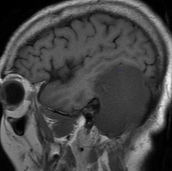
Results: MRI demonstrated a well-limited lesion seen as a hyposignal on T1-weighted images, hypersignal on T2-weighted images, on FLAIR and on diffusion-weighted images not enhanced by gadolinium. The tumour was compressive, and bone lysis was seen on CT.

Conclusion: Epidermoid cysts of the temporal bone are rare, benign lesions whose diagnosis is based on fluid signals seen on MRI but absent on FLAIR sequences.

VIEW THE ENTIRE ARTICLE

References

  • Dietemann J-L. Neuro-imagerie diagnostique. Issy-les-Moulineaux: Elsevier Masson; 2012.
  • Syed MI, Plodpai Y, Khoo SG, Rutka JA. Primary epidermoid cysts of the mastoid: clinical and treatment implications. Eur Arch Otorhinolaryngol 2016;273(4):1055–1059.
  • Twede JV, Patterson MC, Anderson ML. Intraosseous epidermoid cyst of the skull: case study and radiological imaging considerations. Dermatol Online J 2018;24(7):13030/qt5712f7zb.
  • Bikmaz K, Cosar M, Bek S, Gokduman CA, Arslan M, Iplikcioglu AC. Intradiploic epidermoid cysts of the skull: a report of four cases. Clin Neurol Neurosurg 2005;107(3):262–267.
  • Lambert A, Larousserie F, Drapé J-L. Tumeurs osseuses bénignes: kystes épidermoïdes. EMC - Radiologie et Imagerie Médicale - Musculosquelettique - Neurologique - Maxillofaciale 2010;5(4):1–6.
  • Arai A, Sasayama T, Koyama J, Fujita A, Hosoda K, Kohmura E. Epidermoid cyst in Meckel’s cave with unusual computed tomography and magnetic resonance imaging findings. Neurol Med Chir (Tokyo) 2010;50(8):701–704.
  • Saqui AE, Aggouri M, Benzagmout M, Chakour K, Faiz ME. Kyste épidermoïde du quatrième ventricule: à propos d’un cas. Pan Afr Med J 2017;26:239.
  • Views: 502
    HTML downloads: 299
    PDF downloads: 343


    Published: 2020-11-19
    Issue: 2020: Vol 7 No 12 (view)


    How to cite:
    1.
    Abouhanine O, Merzem A, Ndayishimiye V, Belgadir H, Amriss O, Moussali N, El Benna N. Unusual Location of a Recurrent Epidermoid Cyst. EJCRIM 2020;7 doi:10.12890/2020_002009.

    Most read articles by the same author(s)