Masquerading Hypervascular Exophytic Liver Nodule
  • Kevin Kim-Jun Teh
    Department of Gastroenterology and Hepatology, Singapore General Hospital, Singapore
  • Albert Su-Chong Low
    Department of Diagnostic Radiology, Singapore General Hospital, Singapore; Duke-NUS Medical School, Singapore
  • Jason Pik-Eu Chang
    Department of Gastroenterology and Hepatology, Singapore General Hospital, Singapore; Duke-NUS Medical School, Singapore
  • Chee-Kiat Tan
    Department of Gastroenterology and Hepatology, Singapore General Hospital, Singapore; Duke-NUS Medical School, Singapore

Keywords

Exophytic liver nodule, hepatic haemangioma, hepatocellular carcinoma surveillance
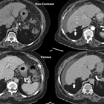
Abstract

Patients with liver cirrhosis are at increased risk of developing hepatocellular carcinoma (HCC) and are placed on routine surveillance for HCC. Diagnosis algorithms are in place to guide clinicians in the evaluation of liver lesions detected during surveillance. Radiological assessments are critical with diagnostic criteria based on identification of typical hallmarks of HCCs on multiphasic computed tomography (CT) and dynamic contrast-enhanced magnetic resonance imaging (MRI). We report a patient with a hypervascular exophytic lesion indeterminate for HCC on CT imaging. While the detection of an exophytic arterially-enhancing lesion in an at-risk patient on CT imaging may prompt clinicians to treat the lesion as HCC without further evaluation, the patient underwent contrast-enhanced MRI with the lesion being eventually diagnosed as an exophytic haemangioma. Thus, no further action was necessary and the patient was continued on routine HCC surveillance.

VIEW THE ENTIRE ARTICLE

References

  • Galle PR, Forner A, Llovet JM, Mazzaferro V, Piscaglia F, Raoul J-L, et al. EASL Clinical Practice Guidelines: management of hepatocellular carcinoma. J Hepatol 2018;69(1):182–236.
  • EASL Clinical Practice Guidelines on the management of benign liver tumours. J Hepatol 2016;65(2):386–398.
  • Cereser L, Furlan A, Bagatto D, Girometti R, Como G, Avellini C, et al. Comparison of portal venous and delayed phases of gadolinium-enhanced magnetic resonance imaging study of cirrhotic liver for the detection of contrast washout of hypervascular hepatocellular carcinoma. J Comput Assist Tomogr 2010;34(5):706–711.
  • Semelka RC, Brown ED, Ascher SM, Patt RH, Bagley AS, Li W, et al. Hepatic hemangiomas: a multi-institutional study of appearance on T2-weighted and serial gadolinium-enhanced gradient-echo MR images. Radiology 1994;192(2):401–406.
  • Views: 791
    HTML downloads: 79
    PDF downloads: 375


    Published: 2020-08-06
    Issue: 2020: Vol 7 No 11 (view)


    How to cite:
    1.
    Teh KK-J, Low AS-C, Chang JP-E, Tan C-K. Masquerading Hypervascular Exophytic Liver Nodule . EJCRIM 2020;7 doi:10.12890/2020_001840.

    Similar Articles

    You may also start an advanced similarity search for this article.

    Most read articles by the same author(s)