Central pontine myelinolysis in a patient undergoing isonatric haemodialysis: the potential role of metabolic and nutritional vulnerability
1 Department of Nephrology and Dialysis, University Clinics of Kinshasa, University of Kinshasa, Kinshasa, Democratic Republic of the Congo
2 Department of Nephrology and Dialysis, University Hospital of French Guiana, Saint-Laurent-du-Maroni Site, French Guiana
3 Department of Nephrology, Dialysis and Kidney Transplantation, University Hospital of Guadeloupe, Guadeloupe
4 Department of Emergency Medicine, University Hospital of French Guiana, Cayenne Site, French Guiana
5 Department of Nephrology and Dialysis, University Hospital of French Guiana, Cayenne Site, French Guiana
Corresponding author’s e-mail: docteur.makarriel2017@gmail.com
Received: 23/10/2026
Accepted: 02/11/2026
Published: 19/11/2026
Conflicts of Interests: The Authors declare no conflict of interest.
Patient Consent: Written informed consent was obtained from the patient for publication of this case report and the accompanying clinical and radiological information, including brain MRI images.
This article is licensed under a CC Attribution-NonCommercial-NoDerivatives 4.0 License
How to cite this article: Makembi Bunkete A, Diallo MM, Muamba Mutombo J, Botaka H, Davodoun T, Djiconkpode I, Gbaguidi T. Central pontine myelinolysis in a patient undergoing isonatric haemodialysis: the potential role of metabolic and nutritional vulnerability. EJCRIM 2026;13:doi:10.12890/2026_007628
ABSTRACT
Introduction: Central pontine myelinolysis (CPM) is usually associated with rapid correction of hyponatremia but may occur in normonatremic patients. Its occurrence during isonatric haemodialysis raises questions about metabolic vulnerability.
Case description: A 65-year-old man receiving maintenance haemodialysis since 2020, subsequently managed with an isonatric strategy, developed progressive tremors, dizziness, gait instability, falls, and cognitive impairment. Brain magnetic resonance imaging in August 2023 revealed a central pontine lesion suggestive of CPM, which was unchanged in April 2024. Previous imaging during an ischemic stroke in 2021 showed no pontine lesion. Predialysis sodium ranged from 138 to 143 mmol/l, without documented rapid correction. Recurrent postdialysis hypokalemia (minimum 3.1 mmol/l), hypoalbuminemia (23 g/l), low prealbumin (12 mg/dl), and inflammation (C-reactive protein 153.4 mg/l) were documented. In June 2024, propranolol, potassium supplementation, and intradialytic parenteral and oral nutritional support were initiated. In July, albumin reached 37.7 g/l, prealbumin 23.6 mg/dl, and C-reactive protein 4.9 mg/l. Treatment was well tolerated. Tremors improved, and the patient regained independence.
Discussion: Recurrent hypokalemia and nutritional and inflammatory abnormalities may increase susceptibility to osmotic demyelination despite limited sodium fluctuations. However, their causal contribution remains uncertain.
Conclusion: CPM should be considered in haemodialysis patients with progressive neurological symptoms, even without documented hyponatremia.
KEYWORDS
Central pontine myelinolysis, hypokalemia, isonatric haemodialysis, malnutrition, osmotic demyelination syndrome
LEARNING POINTS
- Central pontine myelinolysis may occur in patients receiving maintenance isonatric haemodialysis, even without documented hyponatremia or rapid sodium correction.
- Recurrent postdialysis hypokalemia, nutritional impairment, and systemic inflammation may contribute to susceptibility to osmotic demyelination.
- Progressive neurological symptoms in haemodialysis patients warrant careful neurological assessment and evaluation of metabolic and nutritional abnormalities, even when serum sodium remains within the normal range.
INTRODUCTION
Osmotic demyelination syndrome (ODS) is a rare neurological disorder primarily affecting the central pons, classically associated with rapid correction of chronic hyponatremia[1,2]. However, cases occurring in normonatremic patients suggest that other metabolic disturbances and predisposing conditions may contribute to its development[3,4].
Haemodialysis induces changes in circulating solutes, particularly urea, potentially generating osmotic gradients between plasma and cerebral tissues. Although uraemia has been proposed to protect against osmotic demyelination, cases have been reported following haemodialysis despite normal serum sodium concentrations[5,6]. Hypokalemia and malnutrition have also been identified as potential predisposing factors[3,7,8].
Isonatric haemodialysis aims to minimize sodium gradients by individualizing dialysate sodium concentration according to the patient’s average predialysis serum sodium. Nevertheless, this strategy does not eliminate other osmotic changes or underlying metabolic vulnerability.
We report a case of central pontine myelinolysis(CPM) in a patient receiving maintenance isonatric haemodialysis without documented hyponatremia, associated with recurrent postdialysis hypokalemia and marked nutritional and inflammatory abnormalities.
CASE DESCRIPTION
A 65-year-old man with end-stage kidney disease secondary to diabetic nephropathy, receiving maintenance hemodialysis since January 2020, developed progressive tremors, dizziness, gait instability, recurrent falls, and cognitive impairment. His medical history included type 2 diabetes, hypertension, longstanding diabetic peripheral neuropathy, chronic calcific pancreatitis, nutritional impairment, and an ischemic stroke in November 2021 without residual motor deficits. Brain imaging performed at the time of the stroke showed no pontine lesion.
The patient underwent hemodialysis three times weekly for 3.5 hours through a left radiocephalic arteriovenous fistula. An isonatric strategy was introduced in September 2021, with dialysate sodium individualized according to the mean predialysis serum sodium over the preceding three months. The prescribed dialysate contained sodium 140 mmol/l, potassium 3 mmol/l, calcium 1.5 mmol/l, bicarbonate 35 mmol/l, and glucose 1 g/l. Dry weight ranged from 50 to 52.5 kg.
Between January and July 2023, predialysis sodium ranged from 138 to 143 mmol/l, with paired pre- and postdialysis variations between −4 and +1 mmol/l. Urea reduction ratios ranged from 65% to 76%. Recurrent postdialysis hypokalemia (minimum 3.1 mmol/l), hypoalbuminemia (23–31 g/l), low prealbumin (12 mg/dl), and systemic inflammation (C-reactive protein 153.4 mg/l) were documented.
Neurological manifestations progressively worsened during 2022–2023, including disabling hand tremors, impaired balance, recurrent falls, memory difficulties, disorientation, and word-finding difficulties. The patient progressively lost independence, requiring a walking stick indoors and a wheelchair outdoors.
Following treatment, the patient reported a substantial reduction in tremors, which had previously severely impaired his daily activities and independence. He regained autonomy in daily life and described a meaningful improvement in his quality of life.
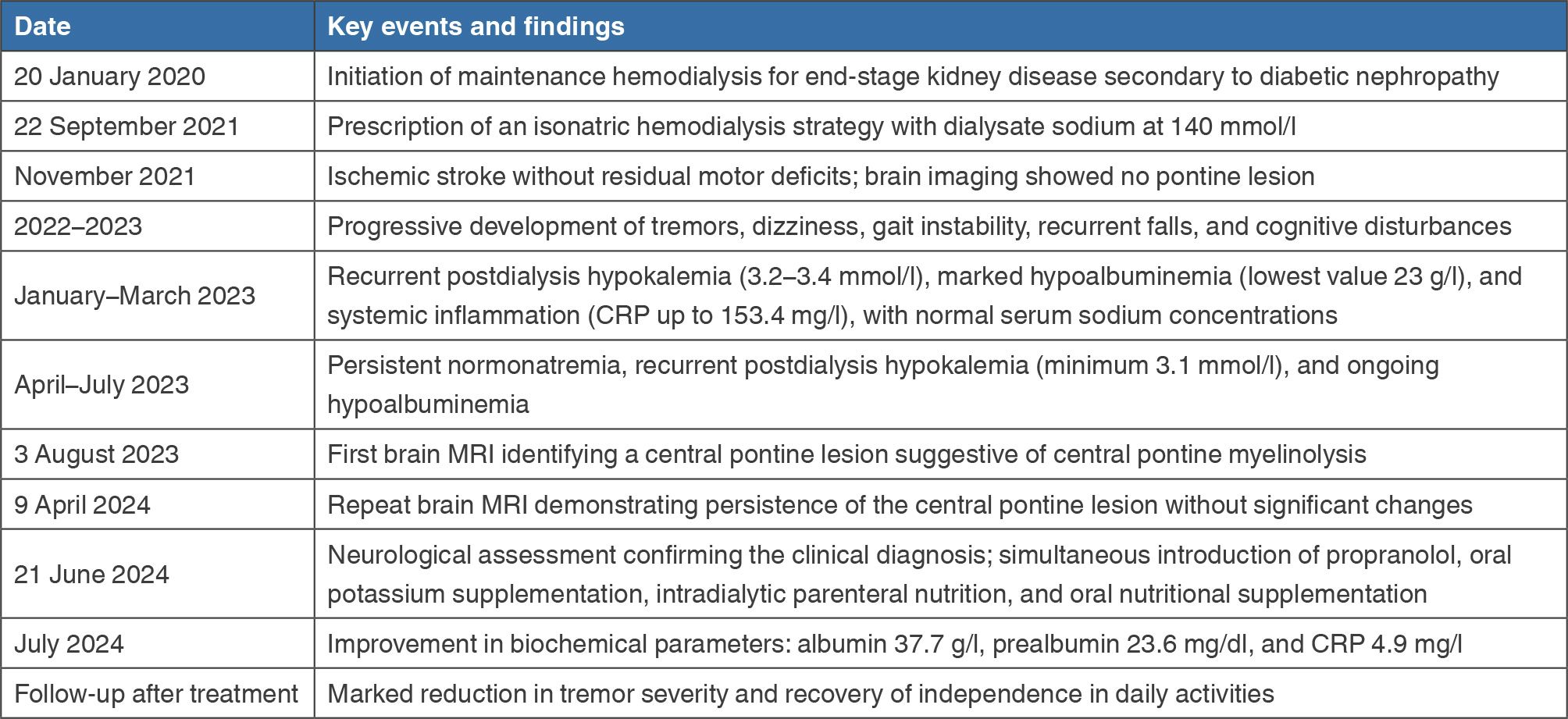
Table 1 summarizes the main clinical events, relevant biochemical abnormalities, diagnostic investigations, and therapeutic interventions.
(click to enlarge)
Table 1. Timeline of major clinical events, relevant laboratory findings, neuroimaging, and treatment.
Abbreviations: MRI, magnetic resonance imaging; CRP, C-reactive protein.
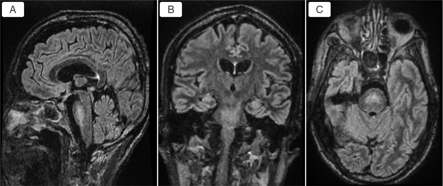
The diagnosis of CPM was based on the progressive neurological manifestations, brain magnetic resonance imaging (MRI) findings, and specialized neurological assessment. A brain MRI performed on August 3, 2023, revealed a central pontine lesion characterized by fluid-attenuated inversion recovery (FLAIR) hyperintensity and mild T1 hypointensity, without diffusion restriction or contrast enhancement. The lesion was visible on sagittal, coronal, and axial FLAIR sequences (Fig. 1). A follow-up MRI performed on April 9, 2024, demonstrated persistence of the pontine lesion without significant interval changes. Importantly, brain imaging performed during the ischemic stroke in November 2021 showed no pontine lesion, indicating that the lesion identified in August 2023 had developed subsequently. Furthermore, diabetic peripheral neuropathy was a longstanding condition that preceded the onset of the progressive neurological manifestations. Neurological examination on June 21, 2024, revealed limb dysmetria, horizontal nystagmus, a positive Romberg sign, and bilateral Babinski signs. Based on the clinical presentation, chronological evolution, and MRI findings, the neurologist retained the diagnosis of CPM. Available biochemical assessments showed no documented hyponatremia or rapid sodium correction. Serum sodium levels before dialysis ranged from 138 to 143 mmol/l, whereas recurrent postdialysis hypokalemia, marked hypoalbuminemia, and elevated inflammatory markers were documented.
(click to enlarge)
Figure 1. Brain magnetic resonance imaging demonstrating a central pontine fluid-attenuated inversion recovery hyperintense lesion in three orthogonal planes: A) sagittal; B) coronal; C) axial.
A comprehensive therapeutic approach was initiated on June 21, 2024, following neurological assessment. Management focused on controlling neurological symptoms, correcting recurrent hypokalemia, and improving nutritional status while maintaining regular hemodialysis. Propranolol was initiated for disabling tremors at 40 mg twice daily for 15 days, followed by 80 mg twice daily. Recurrent postdialysis hypokalemia was treated with oral potassium chloride (Diffu-K 600 mg, one capsule twice daily), providing 16 mmol of potassium per day, with regular biochemical monitoring. Given the marked hypoalbuminemia and nutritional impairment, intradialytic parenteral nutrition was introduced using SmofKabiven E, 493 ml per hemodialysis session, for 1 month. Each bag provided approximately 550 kcal, including 25 g of amino acids, 63 g of glucose, and 19 g of lipids, together with electrolytes, notably 15 mmol of potassium. Oral nutritional supplements were also prescribed for use at home. Maintenance hemodialysis was continued three times weekly using the prescribed isonatric strategy, with ongoing monitoring of electrolyte balance, nutritional and inflammatory parameters, and intradialytic hemodynamic tolerance.
Following the initiation of treatment in June 2024, an improvement in nutritional and inflammatory parameters was observed. In July 2024, serum albumin had increased from 23 to 37.7 g/l, prealbumin from 12 to 23.6 mg/dl, and C-reactive protein had decreased from 153.4 to 4.9 mg/l. All treatments were well tolerated, with no reported adverse events. The patient also reported a marked reduction in tremor severity following propranolol treatment. Before treatment, the tremors had become increasingly disabling, resulting in substantial loss of independence and difficulty performing daily activities. Following treatment, the patient regained autonomy in daily life and reported a significant improvement in quality of life.
DISCUSSION
ODS is classically associated with rapid correction of chronic hyponatremia, although cases occurring in normonatremic patients have been reported[1–4]. Our observation illustrates CPM in a patient receiving long-term isonatric hemodialysis, without documented hyponatremia or substantial intradialytic sodium fluctuations.
Hemodialysis induces changes in circulating solutes, particularly urea, potentially generating osmotic gradients between plasma and cerebral tissues. Although uraemia has been proposed to protect against osmotic demyelination, cases have been reported in normonatremic patients undergoing hemodialysis[5,6]. Thus, maintaining sodium balance through an isonatric strategy may not eliminate other dialysis-related osmotic changes.
Recurrent postdialysis hypokalemia represents a potentially important contributing factor. Previous studies have identified hypokalemia as a predisposing condition for osmotic demyelination[7,8]. In our patient, potassium concentrations repeatedly decreased to 3.1 mmol/l. Marked hypoalbuminemia (23 g/l), low prealbumin (12 mg/dl), and systemic inflammation (C-reactive protein 153.4 mg/l) further suggest metabolic and nutritional vulnerability. These abnormalities may have increased susceptibility to osmotic injury, although their individual contributions cannot be established.
The diagnosis was supported by the characteristic central pontine MRI lesion and neurological findings. Importantly, brain imaging performed during the ischemic stroke in November 2021 showed no pontine lesion, whereas the August 2023 MRI demonstrated a new central pontine abnormality that remained stable on follow-up imaging. Furthermore, diabetic peripheral neuropathy had preceded the progressive neurological manifestations, supporting their distinction from the patient’s pre-existing neurological condition. The diagnosis was retained by the neurologist following clinical and radiological assessment.
Treatment initiated in June 2024 combined propranolol, potassium supplementation, and nutritional support. In July 2024, albumin reached 37.7 g/l, prealbumin 23.6 mg/dl, and C-reactive protein decreased to 4.9 mg/l. Tremors improved substantially, allowing recovery of independence. Although the specific contributions of nutritional support and decreasing inflammation cannot be distinguished, the clinical improvement represents a meaningful therapeutic outcome.
This observation points out the possible contribution of metabolic and nutritional vulnerability to pontine demyelination during isonatric hemodialysis. While the precise underlying mechanism remains uncertain, it emphasizes the importance of considering osmotic demyelination in hemodialysis patients with progressive neurological symptoms, even in the absence of documented sodium disturbances.
CONCLUSION
This case highlights CPM occurring during maintenance isonatric hemodialysis without documented hyponatremia or rapid sodium correction. Recurrent postdialysis hypokalemia and nutritional and inflammatory abnormalities may represent additional predisposing factors. Recognition of these abnormalities, together with appropriate neurological assessment and supportive management, is important in hemodialysis patients presenting with progressive neurological symptoms.