Retinal drusen as an extra-renal manifestation of C3 glomerulopathy
1 Department of Ophthalmology, Dokuz Eylül University, İzmir, Turkiye
2 Department of Nephrology, Başkent University, İzmir, Turkiye
3 Department of Pathology, Memorial Healthcare Group, İstanbul, Turkiye
Corresponding author’s e-mail: osman.saatci@gmail.com
Received: 11/10/2026
Accepted: 27/10/2026
Published: 13/11/2026
Conflicts of Interests: The Authors declare no conflict of interest.
Patient Consent: Written informed consent was obtained from the patient for publication of this report and accompanying images.
This article is licensed under a CC Attribution-NonCommercial-NoDerivatives 4.0 License
How to cite this article: Polat G, Ural Fatihoğlu Ö, Kartı Ö, Ayhan Z, Çelik A, Sarıoğlu S, Saatci AO. Retinal drusen as an extra-renal manifestation of C3 glomerulopathy. EJCRIM 2026;13:doi:10.12890/2026_007603
ABSTRACT
Introduction: C3 glomerulopathy (C3G) is a rare complement-mediated renal disorder caused by dysregulation of the alternative complement pathway. Retinal drusen-like deposits may occur as an extra-renal manifestation.
Case description: A 34-year-old woman with a history of childhood-onset nephrotic syndrome was later classified as having C3G. She underwent living-donor kidney transplantation four years earlier due to end-stage renal disease. She was examined as she was experiencing bilateral blurry vision of long duration and left metamorphopsia. Best-corrected visual acuity was 20/20 in the right eye and 20/40 in the left eye. Fundus examination showed widespread bilateral yellowish drusenoid deposits involving the macula and mid-peripheral retina. Spectral-domain optical coherence tomography demonstrated multiple subretinal pigment epithelium elevations and irregularity of the RPE-Bruch’s membrane complex, while fundus autofluorescence showed mixed hyper- and hypoautofluorescent changes. Renal biopsy material demonstrated intense C3c deposition along glomerular capillary walls with negative immunoglobulin and C1q staining.
Discussion: The shared susceptibility of the glomerular filtration barrier and the RPE-Bruch’s membrane complex to complement-mediated injury may explain this renal-retinal association. Retinal manifestations may remain unrecognised for years, even in kidney transplant recipients.
Conclusion: Clinicians caring for patients with longstanding C3G should be aware of possible ocular involvement. Multimodal retinal imaging can disclose retinal lesions and help clinicians to evaluate vision-threatening complications.
KEYWORDS
C3 glomerulopathy, complement dysregulation, drusen, kidney transplantation, multimodal imaging
LEARNING POINTS
- C3 glomerulopathy may be associated with bilateral retinal drusen as an extra-renal manifestation of complement dysregulation.
- Retinal involvement may remain unrecognised for years; visual symptoms in patients with longstanding C3 glomerulopathy, including kidney transplant recipients, should prompt ophthalmic evaluation.
- Multimodal retinal imaging, particularly spectral-domain optical coherence tomography and optical coherence tomography angiography, can demonstrate retinal involvement and help clinicians assess possible neovascular complications non-invasively.
INTRODUCTION
The complement-mediated renal disorder C3 glomerulopathy (C3G) is rare, and is characterised by dysregulation of the alternative pathway and dominant glomerular C3 deposition, encompassing dense deposit disease and C3 glomerulonephritis, which are distinguished principally by the ultrastructural appearance of glomerular deposits[1,2]. Dysregulation may be genetic or acquired, including autoantibodies such as C3 nephritic factor that stabilise the alternative pathway convertase[2]. The retinal pigment epithelium (RPE)-Bruch’s membrane-choriocapillaris complex shares anatomical and functional similarities with the glomerular basement membrane, including limited local complement regulation, and can become an extra-renal site of complement-mediated deposition[3,4]. A comparable mechanism, involving complement and other immune-mediated constituents, has been proposed to contribute to the formation of classic age-related drusen[5]. Resulting drusen-like lesions typically appear earlier in life than age-related drusen, are often bilateral and extend into the retinal periphery, and can progress to pigment epithelial detachment or choroidal neovascularisation[3,6]; their occurrence in a patient under 50 years of age should prompt consideration of an underlying complement-mediated glomerulopathy[6]. With multimodal imaging, particularly spectral-domain optical coherence tomography and optical coherence tomography angiography, these lesions can now be characterised without invasive dye angiography. We report a 34-year-old woman with childhood-onset nephrotic syndrome and prior kidney transplantation, with bilateral drusenoid deposits on ophthalmic evaluation performed for bilateral blurry vision.
CASE DESCRIPTION
A 34-year-old woman was experiencing bilateral blurry vision and metamorphopsia in the left eye for several months. She was diagnosed with nephrotic syndrome at the age of 9 years and underwent renal biopsy. However, the original pathology report and electron microscopy findings were unavailable for the re-evaluation.
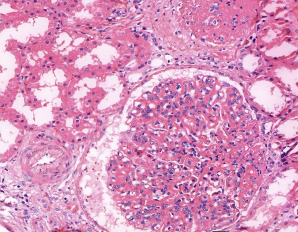
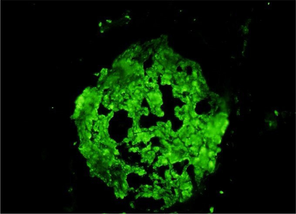
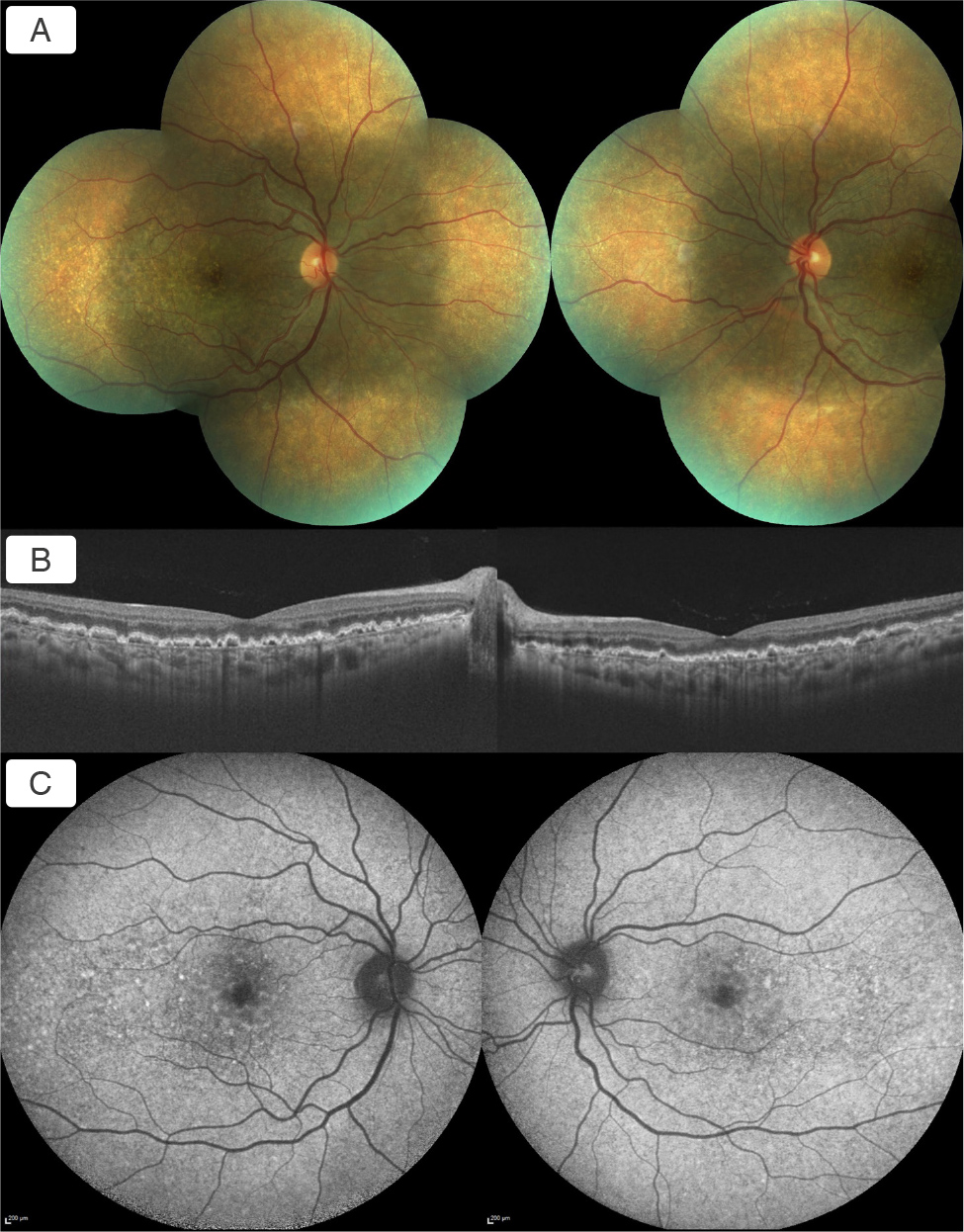
During childhood and adolescence, she received several immunosuppressive regimens including corticosteroids combined sequentially with cyclophosphamide, cyclosporine A and tacrolimus. In 2019, as renal function deteriorated, renal biopsy was repeated, and it was consistent with the initial diagnosis exhibiting predominantly chronic renal parenchymal changes. She subsequently progressed to end-stage renal disease and underwent pre-emptive living-donor kidney transplantation from her mother in 2021. The pre-transplant native kidney biopsy performed in 2019 demonstrated glomerular mesangial expansion, increased cellularity and thickening of the capillary walls (Fig. 1); immunofluorescence showed intense C3c deposition along the glomerular capillary walls and mesangium, with negative staining for IgG, IgA, IgM and C1q (Fig. 2), supporting C3G in the clinical context. According to available medical records, the most recent complement testing, performed in January 2025, showed a markedly reduced serum C3 level of 31 mg/dl (reference range, 90–180 mg/dl); serum C4 was within the normal range at 22 mg/dl (reference range, 10–40 mg/dl). No result for C3 nephritic factor testing was available in the patient’s medical records. At the most recent nephrology evaluation in July 2026, serum creatinine was 1.85 mg/dl, the estimated glomerular filtration rate (eGFR, CKD-EPI) was 34.4 ml/min/1.73 m2, and the spot urine protein-to-creatinine ratio was 781.3 mg/g creatinine. During the patient’s ophthalmological examination in 2025, her best-corrected visual acuity was 20/20 in the right eye and 20/40 in the left eye. Intraocular pressures were normal and anterior segment examination was unremarkable bilaterally. Dilated fundus examination revealed numerous bilateral yellowish drusenoid deposits involving the macula and extending into the mid-peripheral retina (Fig. 3A). Spectral-domain optical coherence tomography demonstrated multiple irregular sub- retinal pigment epithelium (RPE) elevations corresponding to the deposits, with irregularity of the RPE-Bruch’s membrane complex and mild focal attenuation of the overlying outer retinal layers without any intraretinal or subretinal fluid (Fig. 3B). Fundus autofluorescence showed mixed hyper- and hypoautofluorescent changes corresponding to those lesions (Fig. 3C). Optical coherence tomography angiography showed no evidence of macular neovascularisation.
(click to enlarge)
Figure 1. Pre-transplant native kidney biopsy findings. Glomerulus with mesangial expansion, increased cellularity and capillary wall thickening adjacent to a globally sclerosed glomerulus and a thickened arteriole (haematoxylin and eosin, original magnification ×40).
(click to enlarge)
Figure 2. Pre-transplant native kidney biopsy findings. Intense C3c deposition at the mesangium and along the glomerular capillary walls (direct immunofluorescence, C3, original magnification ×40).
(click to enlarge)
Figure 3. A) Colour fundus photographs showing widespread bilateral yellowish drusenoid deposits involving the macula and mid-peripheral retina; B) Spectral-domain optical coherence tomography of both eyes showing multiple sub-RPE drusenoid elevations and irregularity of the RPE-Bruch’s membrane complex, without intraretinal or subretinal fluid; C) Fundus autofluorescence imaging showing a heterogeneous pattern of hyper- and hypoautofluorescence corresponding to the distribution of the drusenoid lesions.
As there was no evidence of exudation or neovascular complication, no ocular treatment was initiated. Regular ophthalmic follow-up with multimodal retinal imaging was advised.
DISCUSSION
The complement-mediated renal disease C3 glomerulopathy is characterised by predominant glomerular C3 deposition and dysregulation of the alternative complement pathway. Dense deposit disease (DDD) and C3 glomerulonephritis lie within this spectrum, with the distinction relying principally on ultrastructural findings[1,2]. Retinal drusen-like deposits are a recognised extra-renal manifestation and were the most frequently reported retinal finding in a recent scoping review of C3 DDD[7], occurring considerably earlier in life than typical age-related drusen[3].
Retinal abnormalities may remain asymptomatic despite the presence of extensive deposits, whereas progressive disease can be associated with retinal pigment epithelial alterations, atrophy or choroidal neovascularisation, the last of which has also been reported in young patients with DDD[3,7,8]. Among 110 native kidney biopsies, seven patients had membranoproliferative glomerulonephritis, including three with biopsy-confirmed DDD. Retinal abnormalities were identified in two patients with DDD, whereas no ocular abnormalities were detected in other membranoproliferative glomerulonephritis subtypes. One patient presented with acute exudative-ischaemic retinopathy showing partial anatomical and functional recovery, while the other exhibited the typical chronic drusenoid phenotype with drusen-like deposits, RPE abnormalities and pigment epithelial detachments[9]. In our patient, widespread bilateral drusenoid deposits involved the macula and mid-peripheral retina. Spectral-domain optical coherence tomography demonstrated sub-RPE elevations and irregularity of the RPE-Bruch’s membrane complex, while optical coherence tomography angiography excluded macular neovascularisation. Similar structural abnormalities have been described with multimodal imaging in C3G[4]. Retinal abnormalities were identified four years after kidney transplantation. Because no ophthalmic examination or retinal imaging was available before transplantation, neither the timing of onset nor the effect of transplantation on the retinal findings can be determined. Previous studies have not established a clear adverse effect of kidney transplantation on the retinal disease[7,10]. Cunningham and Kotagiri[6] reported a 44-year-old patient with retinal pigmentary changes who was followed for over 30 years and underwent renal transplantation with a failed graft a year later. Progressive drusenoid and pigmentary changes developed without any occurrence of choroidal neovascularisation despite renal transplantation, and the authors could not establish a clear relationship between the fundus changes and graft status. It is unclear whether the successful kidney transplantation might have an influence on the retinal findings.
CONCLUSION
Retinal involvement is an important extra-renal manifestation of C3G and may remain clinically unrecognized for years. In patients with longstanding C3G, new visual symptoms should prompt ophthalmic evaluation, while multimodal imaging can help characterize retinal involvement and detect vision-threatening complications. Recognition of this association is particularly relevant in younger patients with atypical drusenoid deposits, in whom an underlying complement-mediated glomerulopathy should be considered. Longitudinal ophthalmic follow-up may help document the evolution of retinal changes and support appropriate long-term clinical surveillance.