Dissociated cholestasis at onset of systemic lupus erythematosus in a male patient: an atypical case
1 Hospital I La Esperanza, EsSalud, Trujillo, La Libertad, Peru
2 Universidad Privada Antenor Orrego, Trujillo, La Libertad, Peru
Corresponding author’s e-mail: jdyf73@gmail.com
Received: 05/10/2026
Accepted: 12/10/2026
Published: 06/11/2026
Conflicts of Interests: The Authors declare no conflict of interest.
Patient Consent: Written informed consent for publication was obtained from the patient.
This article is licensed under a CC Attribution-NonCommercial-NoDerivatives 4.0 License
How to cite this article: Yépez-Florián JD, Yépez-Figueroa GE, De La Cruz-Paucar DE. Dissociated cholestasis at onset of systemic lupus erythematosus in a male patient: an atypical case. EJCRIM 2026;13:doi:10.12890/2026_007583
ABSTRACT
Introduction: Biliary involvement in systemic lupus erythematosus is uncommon, and dissociated cholestasis, defined as elevated alkaline phosphatase and gamma-glutamyl transferase with normal bilirubin, has rarely been described.
Case description: A 41-year-old diabetic man without cutaneous manifestations presented with myalgia and progressive, disabling muscle weakness without creatine kinase elevation. On admission, he had dissociated cholestasis with normal transaminases and concurrent glomerular involvement (proteinuria and haematuria); magnetic resonance cholangiopancreatography was normal. Several days later, he developed rapidly progressive polyserositis requiring urgent pericardiotomy. Strongly positive antinuclear and anti-double-stranded DNA antibodies supported the diagnosis of systemic lupus erythematosus. He improved clinically and biochemically with methylprednisolone and hydroxychloroquine, followed by azathioprine at discharge; alkaline phosphatase and proteinuria normalised at six months, while gamma-glutamyl transferase remained elevated.
Discussion: A normal cholangiogram, the acute course and improvement with immunosuppression made primary sclerosing cholangitis unlikely, although its small-duct variant cannot be excluded without liver biopsy, which was not performed. In the literature reviewed, the dissociated pattern in systemic lupus erythematosus has rarely been described.
Conclusion: In this patient, dissociated cholestasis was a presenting manifestation of systemic lupus erythematosus that coincided with renal involvement and preceded severe serositis. It may be an early marker of lupus activity, a hypothesis that requires validation in a larger series.
KEYWORDS
Dissociated cholestasis, systemic lupus erythematosus, alkaline phosphatase, polyserositis
LEARNING POINTS
- Markedly elevated alkaline phosphatase and gamma-glutamyl transferase with normal bilirubin (dissociated cholestasis) may be a presenting manifestation of systemic lupus erythematosus, even in a man without cutaneous involvement who has disabling muscle weakness and no creatine kinase elevation.
- The dissociated pattern has rarely been described in lupus: among the cases reviewed, the closest one, in a young man, proved on biopsy to be small-duct primary sclerosing cholangitis; therefore, a normal magnetic resonance cholangiopancreatography does not rule out this diagnosis.
- Dissociated cholestasis coincided with renal involvement and preceded severe polyserositis requiring urgent pericardiotomy by several days; in a man with suspected lupus, this finding may warrant close monitoring of disease activity.
INTRODUCTION
Systemic lupus erythematosus (SLE) is an autoimmune connective tissue disease that predominantly affects women, with a female-to-male ratio of 9:1[1]. Gastrointestinal (GI) manifestations of SLE are relatively uncommon, with a prevalence of approximately 10%, but can be severe; ascites, enteritis, pancreatitis and hepatitis are the most common[2]. Hepatobiliary involvement is unusual and typically presents with elevated transaminases, alkaline phosphatase (ALP) and gamma-glutamyl transferase (GGT), whereas dissociated cholestasis (DC) has rarely been reported[2,3]. Patients with GI involvement in SLE often have high disease activity with multiorgan involvement, such as nephritis, polyserositis or cytopenias[1-3]. Our patient presented with DC at disease onset, followed by rapidly progressive polyserositis.
CASE DESCRIPTION
A 41-year-old man had a five-year history of poorly controlled type 2 diabetes mellitus and mixed dyslipidaemia, treated with NPH insulin (20 IU at breakfast and 12 IU at dinner), metformin 850 mg at breakfast and lunch, and atorvastatin 20 mg at 9 pm. Laboratory tests before the current illness had shown hyperglycaemia but normal liver enzymes.
The illness began with subjective, unmeasured fever, mild headache and moderate generalised myalgia (VAS 4/10), predominantly in the lower limbs, followed by temporomandibular joint pain with dysphagia. Myalgia then worsened and joint pain extended to the shoulders and knees. By around day 9, proximal lower-limb weakness, odynophagia and otalgia had developed. As proximal and distal weakness progressed to partial immobility, the patient was hospitalised on illness day 13.
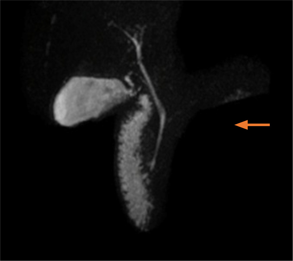
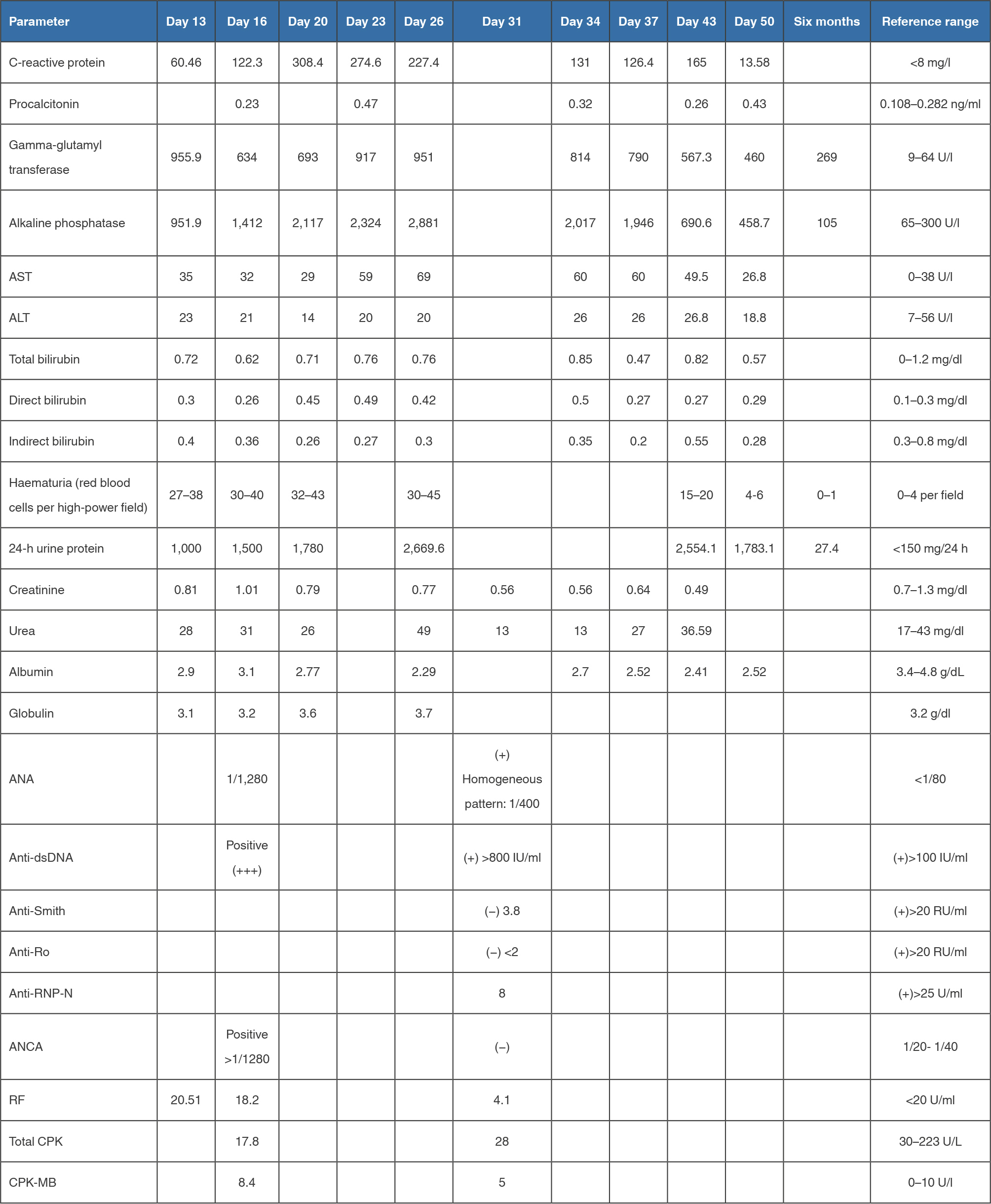
Blood tests showed elevated ALP (951.9 U/l) and GGT (955.9 U/l) without bilirubin or transaminase abnormalities, consistent with a DC pattern (Table 1). Glomerular involvement with proteinuria and haematuria was documented, and positive antinuclear antibodies (ANA) and antineutrophil cytoplasmic antibodies (ANCA) raised suspicion of autoimmune vasculitis. Abdominal ultrasound ruled out obstructive biliary disease. The differential diagnoses considered were polymyositis, SLE, systemic vasculitis and overlap syndrome with primary sclerosing cholangitis (PSC). On day 16, he developed fever without leucocytosis, elevated C-reactive protein and an inverted albumin-to-globulin ratio, with normal procalcitonin and non-elevated creatine kinase. Dyspnoea then progressed until it was present at rest, and he was transferred to the intensive care unit (ICU). Multislice computed tomography (MSCT) of the chest confirmed serositis with pleural and pericardial effusion; 36 hours after ICU admission, follow-up MSCT showed an increase in the pericardial effusion, left basal consolidation and mild ascites. Initial serology obtained on day 16, at ICU transfer, showed ANA 1/1280 and strongly positive anti-double-stranded DNA (anti-dsDNA); a confirmatory immunological panel on day 31 showed positive ANA (homogeneous pattern, 1/400) and anti-dsDNA >800 IU/ml, with negative anti-RNP-N and ANCA (Table 1). Magnetic resonance cholangiopancreatography (MRCP) showed no biliary tree abnormalities, excluding obstructive biliary disease (Fig. 1). Liver biopsy was deferred at the request of the patient and his family and was ultimately not performed in view of the progressive clinical and biochemical improvement.
(click to enlarge)
Figure 1. Magnetic resonance cholangiopancreatography. No structural abnormality is identified (arrow).
In the ICU, he received high-flow oxygen therapy and was started on meropenem and clindamycin. On the third ICU day, mixed connective tissue disease (MCTD) was suspected and prednisone 10 mg/day was started. Because of further clinical deterioration, urgent pericardiotomy was performed, draining 250 ml of cloudy fluid with a cell count of 3,283 cells/µl, predominantly polymorphonuclear (97%), and the antibiotic regimen was changed to meropenem and vancomycin. As oxygen requirements decreased, the patient was transferred to the internal medicine ward and started on intravenous methylprednisolone 500 mg/day for three days, followed by tapering prednisone and hydroxychloroquine 400 mg/day, with mycophenolate 500 mg every 12 hours planned after completion of the antibiotic course. He remained transiently dependent on supplemental oxygen. C-reactive protein rose again to 165 mg/l on day 43, when a new course of meropenem was started, and subsequently fell to 13.58 mg/l at discharge.
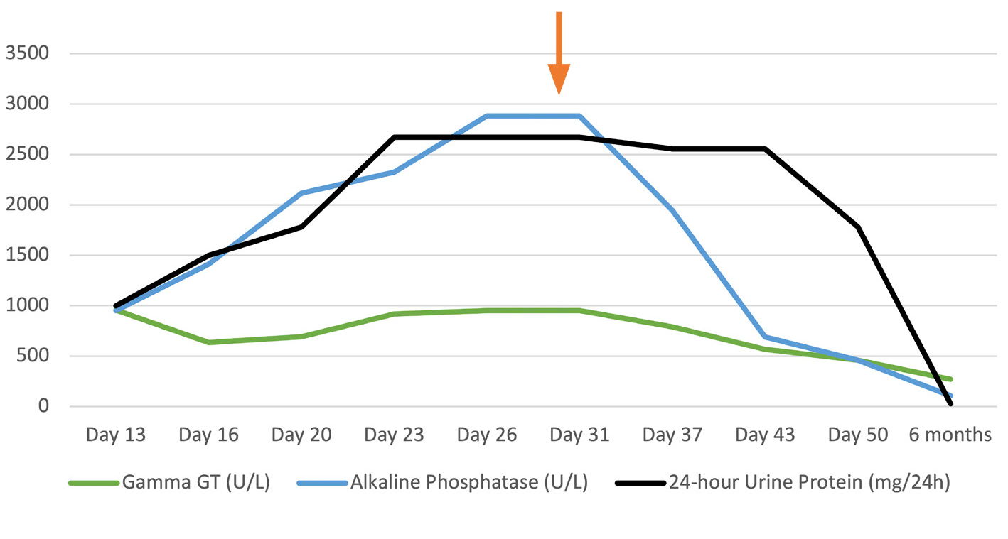
He was discharged on day 50, after marked decreases in ALP, GGT and proteinuria, together with reductions in C-reactive protein and haematuria. The discharge regimen included prednisone (20 mg at 8 am and 10 mg at 4 pm), azathioprine 50 mg every 12 hours and hydroxychloroquine 400 mg every 24 hours. By 6 months of outpatient follow-up, proteinuria and ALP had normalised (Fig. 2), whereas GGT had decreased but remained above the reference range (269 U/l; Table 1).
(click to enlarge)
Figure 2. Temporal correlation between the decrease in cholestatic enzymes and proteinuria, the days of illness and the start of corticosteroid therapy (arrow). Note: ICU admission on day 16; methylprednisolone pulse started on day 31.
(click to enlarge)
Table 1. Blood tests.
DISCUSSION
The DC pattern is characterised by elevated GGT and ALP, with preserved bilirubin excretion and normal serum bilirubin levels[4], a profile consistent with that observed in our patient. The Systemic Lupus Erythematosus Disease Activity Index (SLEDAI) does not include GI manifestations, and liver dysfunction is not included in the classification criteria of the disease[5]. DC may be intrinsic to SLE or indicate an overlap syndrome; establishing its aetiology is therefore essential for appropriate management[3].
This case describes the onset of lupus in an adult man without cutaneous manifestations, with myalgia and disabling progressive muscle weakness. The normal MRCP, the acute course and the biochemical improvement under immunosuppression made PSC (which predominates in men) unlikely, although its small-duct variant, defined by normal cholangiography, cannot be excluded without histology[6,7]. Non-elevated creatine kinase and negative anti-RNP-N made polymyositis and MCTD unlikely, and the subsequent negative ANCA result weighed against systemic vasculitis. DC was therefore attributed to SLE, supported by positive ANA and anti-dsDNA (Table 1). Compared with women, men with SLE more frequently have renal manifestations and accrue more neuropsychiatric, renal and cardiovascular damage[8]. In our patient, renal involvement coincided with DC, which preceded polyserositis by several days.
In the literature reviewed, Chow et al.[9] described an icteric pattern, also at SLE onset, with a total bilirubin of 147 µmol/l. Oh et al.[7] described a dissociated pattern (ALP 860 U/l, GGT 802 U/l and normal bilirubin) in a 24-year-old man; however, biopsy established small-duct PSC. Histology was available in both cases. To our knowledge, the combination of a dissociated pattern, male gender, presentation at disease onset and biochemical improvement without histology has not been described in the literature reviewed.
Corticosteroids are the mainstay of treatment for acute GI involvement in SLE, with some evidence supporting their combination with immunomodulatory agents[2]. In our patient, this strategy (methylprednisolone, hydroxychloroquine and azathioprine) was associated with clinical and biochemical improvement and a progressive decrease in systemic activity, including DC (Fig. 2). This finding is compatible with the diagnosis, although the response to corticosteroids is non-specific and does not replace histological confirmation.
Azathioprine, started at discharge, is a recognised cause of cholestatic liver injury including ductopenia, in patients with SLE[10]. The persistently elevated GGT at 6 months could suggest a drug contribution; however, this is unlikely because cholestasis preceded azathioprine and ALP normalised during its use.
For a single case, this study cannot establish causality or estimate predictive value. The temporal sequence of DC, renal involvement and polyserositis does not exclude contributing factors such as poorly controlled diabetes, infection treated with broad-spectrum antibiotics or drug exposure, which are recognised causes of elevated ALP and GGT. Liver biopsy was not performed and antimitochondrial antibodies were not measured; nevertheless, primary biliary cholangitis is unlikely given its female predominance, and the normal MRCP excluded large-duct PSC[6]. Because GGT remained elevated at six months, longer follow-up is required to confirm complete biochemical resolution.
CONCLUSION
We report the onset of severe SLE in a man whose atypical presentation was characterised by the initial coexistence of renal involvement and DC, which preceded rapidly progressive polyserositis. The temporal sequence observed suggests that DC may serve as an early marker of severe lupus activity, a hypothesis that a single case cannot confirm. Analytical studies are needed to determine whether biliary involvement adds information to SLE activity indices.