Tertiary hyperparathyroidism in a patient with autosomal dominant pseudohypoparathyroidism type 1b
Department of Endocrinology, Epicura Hospital, route de Mons 63, Hornu, Belgium
Corresponding author’s e-mail: vandemergel@hotmail.fr
Received: 02/10/2026
Accepted: 12/10/2026
Published: 20/11/2026
Conflicts of Interests: The Author declares no conflict of interest.
Patient Consent: Informed consent was obtained from the patient for the publication of this case data.
This article is licensed under a CC Attribution-NonCommercial-NoDerivatives 4.0 License
How to cite this article: Vandemergel X. Tertiary hyperparathyroidism in a patient with autosomal dominant pseudohypoparathyroidism Type 1b. EJCRIM 2026;13:doi:10.12890/2026_007581
ABSTRACT
Background: Pseudohypoparathyroidism type 1b (PHP1b) is a rare genetic disorder characterized by renal parathyroid hormone (PTH) resistance. While chronic secondary hyperparathyroidism is common, progression to autonomous tertiary hyperparathyroidism remains exceptionally rare, with only five cases reported in the literature.
Case description: We report the case of a 66-year-old Caucasian woman with an autosomal dominant form of PHP1b (maternal transmission of a STX16 gene deletion) diagnosed at age 35. After 31 years of stable disease managed with calcitriol, she presented with persistent hypercalcemia for 5 years. Longitudinal follow-up revealed a dramatic, tenfold surge in serum intact PTH levels, peaking at 932 ng/l. Physical examination showed a mildly rounded facies and brain imaging revealed basal ganglia calcifications. A neck ultrasound and technetium-99m sestamibi scintigraphy confirmed a right medial parathyroid nodule. The patient underwent surgical resection of the lesion. Histopathology confirmed a follicular parathyroid adenoma. Postoperatively, serum calcium and PTH levels normalized rapidly and remained stable at 6-month follow-up.
Conclusion: Tertiary hyperparathyroidism is a rare but critical long-term complication of PHP1b that typically occurs after decades of stimulation. In patients with PHP1b, the onset of hypercalcemia accompanied by a significant, progressive increase in PTH levels should immediately raise suspicion of an autonomous parathyroid adenoma and prompt targeted imaging and surgical management.
KEYWORDS
Pseudohypoparathyroidism type 1b (PHP1b), STX16 deletion, tertiary hyperparathyroidism, parathyroid adenoma
LEARNING POINTS
- Pseudohypoparathyroidism is a rare disease rarely complicated by tertiary hyperparathyroidism.
- A change in phosphate-calcium metabolism with hypercalcemia should alert the clinician.
INTRODUCTION
Pseudohypoparathyroidism (PHP) is a group of rare disorders characterized by parathyroid hormone (PTH) resistance at the renal tubule, associated with a spectrum of physical, neurocognitive, and endocrine abnormalities. Primarily caused by molecular defects impairing hormone signalling via receptors coupled through the α-subunit of the stimulatory G protein (Gs-α) and adenylyl cyclase[1], its exact prevalence is unknown, but it is estimated to range between 0.34 per 100,000 in Japan and 1.1 per 100,000 in Denmark[2,3]. Several distinct clinical entities have been described, affecting various organs with differing degrees of severity. Patients with PHP1b typically display imprinting or methylation defects in the GNAS gene on chromosome 20, leading to isolated PTH resistance in the absence of Albright hereditary osteodystrophy (AHO) features[1]. However, recent studies have shown that some patients with PHP1b exhibit additional features overlapping with PHP1a. Although patients with chronically elevated PTH are at risk of developing autonomous parathyroid tumors (tertiary hyperparathyroidism), this complication remains exceptionally rare[4]. We report the case of a female patient with a history of PHP1b complicated by tertiary hyperparathyroidism.
CASE DESCRIPTION
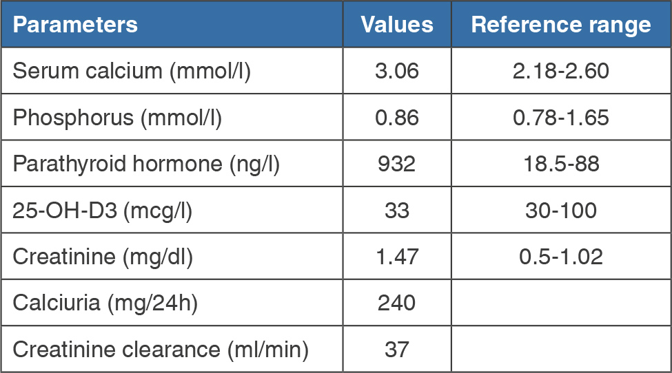
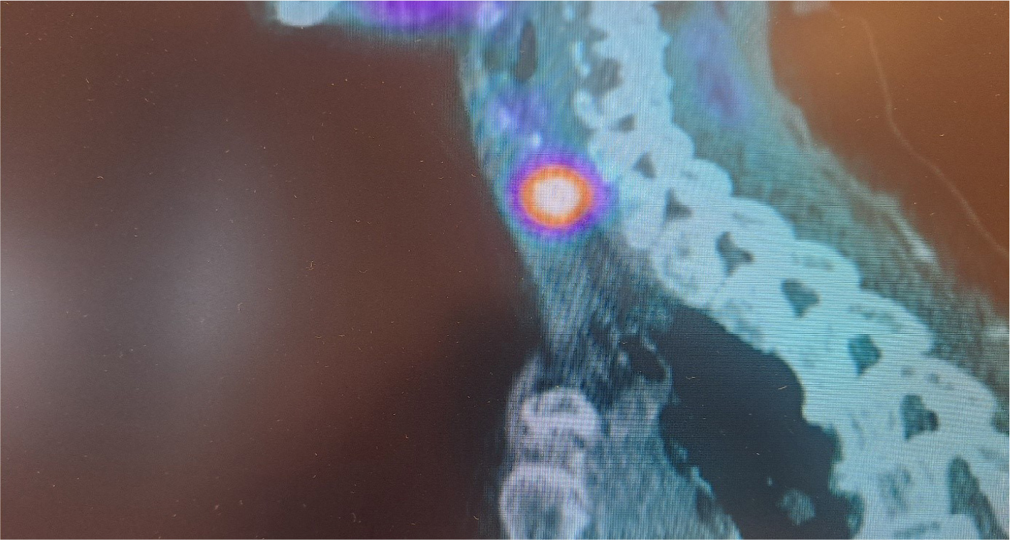
A 66-year-old Caucasian woman consulted the endocrinology department for calcium and phosphate metabolism disorders. She had a history of PHP1b diagnosed at the age of 35. Her daughter and granddaughter also presented with the same condition (and were found to carry an STX16 gene deletion). Her other medical history included previously treated Hashimoto’s thyroiditis, hyperlipidemia, osteoporosis with a T-score of -2.1 at the femoral neck, and -2.6 at the lumbar spine, chronic kidney disease, and lumbar osteoarthritis. She had no history of nephrolithiasis. Her maintenance treatment included levothyroxine and 0.5 µg of calcitriol daily. Physical examination revealed a mildly rounded facies, a weight of 65 kg, and a height of 165 cm; blood pressure was 130/80 mmHg. The physical examination was otherwise normal. Laboratory findings are summarized in Table 1. Neck ultrasound revealed a right medial parathyroid nodule measuring 10 × 7 × 8 mm. A review of the medical records revealed a history of hypercalcemia for five years prior to presentation, which persisted despite discontinuation of calcium supplementation, along with markedly elevated PTH levels. PTH levels ranged between 79 and 142 ng/l until 2018, rising to 393 ng/l and 645 ng/l in 2019 and 2020, respectively. The peak PTH level prior to surgery was 932 ng/l. Given the suspicion of tertiary hyperparathyroidism, the patient underwent sestamibi parathyroid scintigraphy (Fig. 1), which confirmed a right parathyroid adenoma. Of note, previous brain imaging revealed basal ganglia calcifications. Surgical resection was recommended, and histopathology confirmed a follicular parathyroid adenoma. Serum calcium levels normalized rapidly after surgery (2.4 mmol/l) and postoperative PTH levels dropped rapidly to 32 ng/dl. Six months after surgery, serum calcium levels were normalized at 2.15 mmol/l, with the urinary calcium-to-creatinine ratio at 0.12.
(click to enlarge)
Table 1. Laboratory workup at presentation.
(click to enlarge)
Figure 1. Sestamibi scintigraphy showing increased tracer uptake in the right lower neck region consistent with the lesion identified on ultrasound.
DISCUSSION
To our knowledge, only five cases of patients with PHP1b complicated by tertiary hyperparathyroidism have been previously reported in the literature[4]. All reported cases occurred in women. The diagnosis is generally delayed and is prompted by a progressive increase in serum calcium and PTH levels. In this case, although serum calcium levels remained within the normal range during treatment with calcitriol and vitamin D, the onset of hypercalcemia occurred at least five years prior to presentation (compared to a mean of two years in Neary et al.’s study[4]).
The time interval between the diagnosis of PHP1b and the onset of tertiary hyperparathyroidism is usually long. It was 31 years in our patient compared with 34 years in Neary et al.’s series of five cases. This time interval is also extremely long (32 and 25 years) in the rare cases reported in patients with tertiary hyperparathyroidism secondary to PHP1a[5].
It should be noted that PTH monitoring reveals significantly elevated levels when tertiary hyperparathyroidism develops. In this case, the PTH level had more than doubled. In the Neary et al. series, baseline values were not consistently reported, but in the two cases where they were known, the increase was 40% and 66%, respectively. Similarly, a rapid return to baseline values is observed following surgery. Among the four operated patients in the Neary et al. series, two achieved subnormal PTH levels, while the other two showed a decrease of 66% and 95%, respectively.
Theoretically, tertiary hyperparathyroidism could be caused by a single adenoma but could also result from multiple lesions or diffuse hyperplasia. However, in all reported cases, the adenoma was isolated. In two cases, a second parathyroid lesion was resected based on the intraoperative findings, although it was not confirmed whether it was also an adenoma.
Clinically, patients with PHP1b typically do not present with the clinical features of AHO. Furthermore, cerebral calcifications, which are common to both PHP1a and PHP1b, are found in up to 70% of cases[6]. These calcifications are found primarily in the basal ganglia but may extend to the thalamus and the cortex[1].
Although resistance to thyroid-stimulating hormone cannot be excluded, the thyroid dysfunction observed in this case appears to be related to a history of Hashimoto’s thyroiditis.
This case is also unique in that it is inherited in an autosomal dominant manner, a mode of inheritance that accounts for only 15-20% of cases, while the remaining cases are sporadic[7].
CONCLUSION
Although rare, tertiary hyperparathyroidism can develop as a serious complication in patients with PHP1b. The onset of hypercalcemia accompanied by elevated PTH levels should prompt the clinician to investigate for a parathyroid adenoma.