Diagnostically challenging Pro-B ALL followed by emergence of monocytic AML after successful CAR-T treatment
1 The Cellular Immunophenotyping Unit, Hematology Institute, Tel Aviv Sourasky Medical Center, Tel Aviv, Israel
2 Gray Faculty of Medical and Health Sciences, Tel Aviv University, Tel Aviv, Israel
Corresponding author’s e-mail: chensa@tlvmc.gov.il
Received: 26/08/2026
Accepted: 07/09/2026
Published: 06/11/2026
Conflicts of Interests: The Authors declare no conflict of interest.
Patient Consent: Written informed consent waived by the Tel Aviv Medical Center IRB committee.
This article is licensed under a CC Attribution-NonCommercial-NoDerivatives 4.0 License
How to cite this article: Glait-Santar C, Katz BZ. Diagnostically challenging Pro-B ALL followed by emergence of monocytic AML after successful CAR-T treatment. EJCRIM 2026;13:doi:10.12890/2026_007474
ABSTRACT
Introduction: Therapy-related acute myeloid leukemia (t-AML) is a recognized complication of leukemia-directed cytotoxic therapy. However, the emergence of therapy-related monocytic AML shortly after successful CD19-directed chimeric antigen receptor T-cell (CAR-T) therapy for relapsed Pro-B acute lymphoblastic leukemia (B-ALL) raises important questions regarding clonal evolution, lineage plasticity, and treatment-related leukemogenesis.
Case Description: We describe a 73-year-old man diagnosed with Philadelphia chromosome-negative Pro-B ALL with hyperdiploidy/tetraploidy, whose initial presentation posed a diagnostic challenge due to expression of immature and partial myeloid-associated markers, initially suggesting minimally differentiated AML. Repeat immunophenotyping clarified the diagnosis as Pro-B ALL. The patient achieved complete remission with the GMALL Elderly protocol despite severe infectious and treatment-related complications. Following relapse during maintenance therapy, he received salvage treatment with inotuzumab ozogamicin followed by CD19-directed CAR-T therapy, achieving marrow remission. Five months later, he developed secondary monocytic AML with no immunophenotypic evidence of residual B-ALL.
Conclusion: This case underscores the importance of precise immunophenotypic characterization in diagnostically ambiguous acute leukemias and illustrates the complex biologic evolution that may follow sequential chemotherapy and modern immunotherapeutic approaches, including the emergence of secondary myeloid neoplasms after CAR-T therapy.
KEYWORDS
Acute leukemia, monocytic leukemia, myeloid neoplasm, multiparameter flow cytometry
LEARNING POINTS
- Extended flow cytometric immunophenotyping is essential to correctly distinguish minimally differentiated acute myeloid leukemia (AML) from Pro-B acute lymphoblastic leukemia (ALL) when initial blast marker expression is ambiguous.
- Sequential exposure to chemotherapy, inotuzumab ozogamicin, and CD19-directed chimeric antigen receptor t-cell therapy may be followed by a distinct secondary monocytic AML, independent of the original B-ALL clone.
- Repeat bone marrow evaluation with comprehensive immunophenotyping is needed at relapse or disease evolution to distinguish secondary AML from relapsed/lineage-switched B-ALL and guide therapy.
INTRODUCTION
Acute lymphoblastic leukemia (ALL) in elderly patients remains associated with substantial morbidity and mortality owing to adverse disease biology, frailty, infectious complications, and limited tolerance to intensive therapy[1,2]. Nevertheless, incorporation of modern treatment protocols and targeted immunotherapies has improved remission rates and survival outcomes in selected patients[3]. Secondary myeloid neoplasms, including secondary AML, are well-recognized complications of cytotoxic chemotherapy and radiation exposure accounting for 10-20% of all AML cases and occurring in 1-10% of cancer survivors over approximately 10 years[4]. Increasing use of targeted immunotherapies such as inotuzumab, ozogamicin and CD19-directed chimeric antigen receptor T-cell (CAR-T) therapy has introduced additional selective pressures whose long-term hematopoietic effects remain incompletely understood[5]. Distinguishing recurrent ALL from secondary AML is critical because these entities require fundamentally different therapeutic approaches and carry distinct prognostic implications. In our case, the diagnosis becomes particularly challenging when patients present with ambiguous immunophenotypic features followed by secondary monocytic AML[6].
Such distinction is critical because therapeutic strategies, prognosis, and biologic implications differ substantially[7,8]. We report a diagnostically interesting case of Philadelphia chromosome-negative Pro-B ALL with hyperdiploidy/tetraploidy in an elderly patient who achieved remission with GMALL Elderly therapy, relapsed during maintenance therapy, underwent salvage treatment with inotuzumab ozogamicin followed by CD19-directed CAR-T therapy, and subsequently developed secondary monocytic AML without evidence of residual lymphoblastic leukemia.
CASE DESCRIPTION
A 73-year-old previously independent man was admitted in April 2022 because of febrile neutropenia and pancytopenia. His medical history included chronic pansinusitis status post functional endoscopic sinus surgery in 2019, benign prostatic hyperplasia, and ischemic heart disease treated with coronary catheterization earlier in 2022. He had no smoking history and no history of alcohol or illicit drug use.
Initial laboratory studies demonstrated pancytopenia with hemoglobin levels of 9-10 g/dl, white blood cell count of 0.5×10⁹/l, profound neutropenia, platelet count around 50×109/l, and markedly elevated lactate dehydrogenase levels (>1000 µ/l).
Bone marrow examination performed at the referring institution demonstrated marrow replacement by approximately 40% blasts lacking granulation or Auer rods. Initial flow cytometric analysis revealed positivity for HLA-DR, CD4, CD13, CD34, CD117, CD38, CD123, and TdT, while MPO and multiple lymphoid markers were negative. As part of the diagnostic evaluation, conventional Fluorescence in situ hybridization studies (FISH) analysis demonstrated a male hyper diploid, tetraploid karyotype. FISH studies showed no evidence of KMT2A rearrangement, t(8;21) or inv(16). Molecular testing was negative for NPM1 mutation, FLT3-ITD mutation, and BCR::ABL1 transcripts. Based on these findings, the patient was initially diagnosed with minimally differentiated AML (AML-M0).
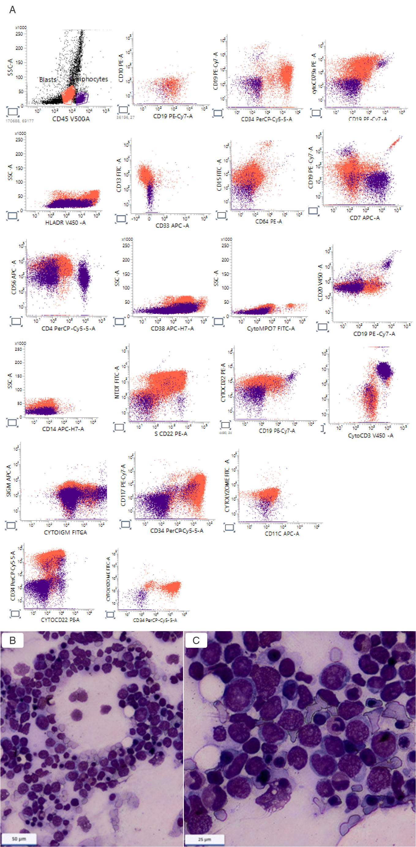
Following transfer to our institution, bone marrow aspiration demonstrated approximately 40% blasts with moderate erythroid dysplasia. Flow cytometric analysis demonstrated positivity for CD22, TdT, CytoCD79a, CD45, CD117, HLA-DR, CD13, cytoplasmic IgM and CD34. Cytoplasmic MPO, lysozyme, CD64, CD14, CD33, CD56, CD3, CD7, CD19, CD10, CD20, and surface IgM were negative (Fig. 1A). Bone marrow infiltrated by monomorphic lymphoblasts with high nuclear-to-cytoplasmic ratios, fine chromatin, inconspicuous nucleoli, and scant agranular basophilic cytoplasm (Fig. 1B and 1C).
(click to enlarge)
Figure 1. A) Multiparameter flow cytometric immunophenotyping demonstrating B-lineage acute lymphoblastic leukemia (B-ALL); B, C) Wright–Giemsa–stained bone marrow aspirate smears at diagnosis of pro-B ALL to acute monocytic acute myeloid leukemia.
Conventional FISH analysis demonstrated a male hyper diploid, tetraploid karyotype, without chromosomal 5 and 7 aberrations and no evidence for translocations involving MLL gene or 8:21. FISH studies showed no evidence of KMT2A rearrangement, t(8;21) or inv(16). Molecular testing was negative for NPM1 mutation, FLT3-ITD mutation, and BCR::ABL1 transcripts. Molecular analysis by real-time PCR at diagnosis showed no evidence of the t(4;11) translocation (KMT2A), and testing for the BCR::ABL1 p210 and p190 fusion transcripts were negative, consistent with Philadelphia chromosome-negative disease. Integration of morphologic, immunophenotypic, molecular, and cytogenetic findings established the diagnosis of Philadelphia chromosome-negative Pro-B ALL with hyper diploidy/tetraploid. Lumbar puncture demonstrated no evidence of central nervous system involvement. Computed tomography imaging demonstrated mild splenomegaly containing nonspecific hypodense lesions and borderline enlarged porta hepatis lymph nodes.
Induction treatment according to the GMALL Elderly protocol was initiated on May 3, 2022. Therapy included dexamethasone followed by cyclophosphamide, vincristine, idarubicin, dexamethasone, and intrathecal methotrexate/cytarabine/dexamethasone. Day 15 treatment was omitted because of prolonged aplasia and infectious complications.
Hospitalization was complicated by profound treatment-related immunosuppression with Pseudomonas aeruginosa bacteremia, cellulitis, and severe Pneumocystis jirovecii pneumonia (confirmed by bronchoalveolar lavage polymerase chain reaction), requiring prolonged antimicrobials, trimethoprim-sulfamethoxazole, corticosteroids, and respiratory support. Additional toxicities included fluid retention, generalized weakness, and functional decline requiring physiotherapy. Despite these complications, bone marrow examination by flow cytometric analysis performed on day 20 of induction (May 23, 2022) demonstrated complete remission. The patient subsequently continued treatment according to the GMALL Elderly protocol and remained in remission. By March 2023, during week 41 of therapy, blood counts had significantly improved with hemoglobin of 11.7 g/dl and platelet count of 112×109/l. In June 2023, maintenance therapy with mercaptopurine and methotrexate was initiated.
In January 2024, progressive thrombocytopenia and leukopenia developed, accompanied by relative eosinophilia and basophilia without circulating blasts. Lactate dehydrogenase, bilirubin, and renal function remained normal. A month later, relapse of B-ALL was confirmed by multicolor flow cytometry during maintenance therapy. The patient subsequently underwent leukapheresis and received salvage treatment with inotuzumab ozogamicin as bridging therapy followed by CD19-directed CAR-T therapy on March 24, 2024. A month later, there was no immunophenotypic evidence of residual B-ALL by flow cytometry. The post-CAR-T course was complicated by grade 2 cytokine release syndrome, grade 2 immune effector cell-associated neurotoxicity syndrome, neutropenic fever, vancomycin-resistant Enterococcus colonization, Clostridioides difficile-associated diarrhea, coagulopathy, anorexia, weight loss, intermittent confusion, and marked functional deterioration.
Five months after relapsed Pro-B ALL, the patient was hospitalized with leukocytosis, thrombocytopenia, fever, acute kidney injury, hyperuricemia, elevated lactate dehydrogenase, and hypophosphatemia. Bone marrow examination performed and demonstrated approximately 70% blasts with monocytic morphology and immunophenotypic features consistent with therapy-related AML, without evidence of residual B-ALL detected by flow cytometry.
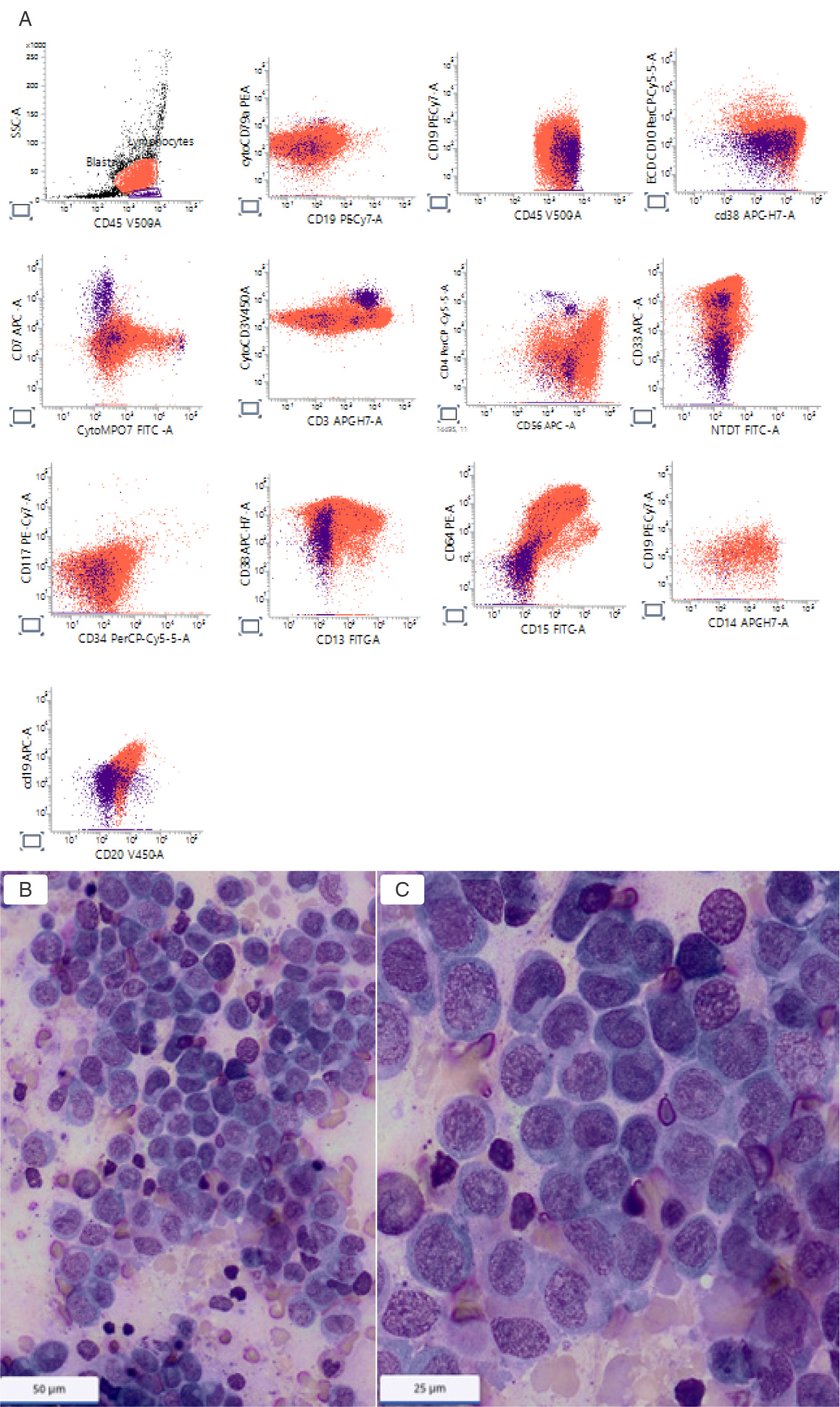
At that time, treatment with azacitidine and venetoclax was initiated. The clinical course was complicated by spontaneous tumor lysis syndrome with worsening renal dysfunction, electrolyte abnormalities, metabolic acidosis, and subsequent transient improvement following aggressive supportive management. Bone marrow examination on day 21 demonstrated persistent active leukemia with approximately 30% blasts by flow cytometry (Fig. 2A). Infiltration of the bone marrow by monomorphic monoblasts with irregular/folded nuclei, delicate chromatin, and prominent nucleoli, illustrate the distinctly different morphology of the subsequent secondary neoplasm (Fig. 2B and 2C). During hospitalization, the patient sustained a fall complicated by subdural hemorrhage and vertebral compression fractures. Progressive refractory leukemia, worsening performance status, and continued clinical deterioration ultimately culminated in death in July 2024.
(click to enlarge)
Figure 2. A) Flow cytometry histograms of the bone marrow aspirate demonstrating a distinct monocytic population (red); B, C) Wright–Giemsa–stained bone marrow aspirate smears at diagnosis of secondary monocytic acute myeloid leukemia.
DISCUSSION
This case highlights key diagnostic and biologic challenges in modern leukemia management. The initial immunophenotypic profile was ambiguous: expression of immature markers and partial myeloid antigens (CD13, CD117) with absent classic B-lineage markers (CD19, CD10) initially suggested AML-M0. Repeat evaluation, however, demonstrated cytoplasmic IgM, CD22, CD79a, and TdT, confirming Pro-B ALL[6]. The expression of CD19 plus cytoplasmic CD79a and/or cytoplasmic CD22 are essential for B-ALL diagnosis[6]. Cytoplasmic IgM supports pre-B cell stage differentiation. The case described illustrates a well-documented diagnostic pitfall where initial expression of CD13 and CD117 with absent classic B-lineage surface markers can wrongly be diagnosed as AML-M0, but subsequent detection of cytoplasmic IgM, CD22, CD79a, and TdT correctly reclassified the case as Pro-B ALL[9].
This distinction carried major therapeutic significance[7,8]. The case underscores the critical importance of comprehensive immunophenotypic analysis, integration of morphology and molecular findings, and repeat bone marrow assessment in diagnostically ambiguous acute leukemias. From a laboratory perspective, this case highlights how incomplete lineage assignment may lead to fundamentally different therapeutic pathways and prognostic implications.
Despite advanced age and profound induction-related toxicity—including aplasia, Pseudomonas bacteremia, and severe Pneumocystis jirovecii pneumonia—he achieved complete remission, sustained for nearly two years. The later disease course reflects the evolving therapeutic landscape of relapsed B-ALL. Following relapse during maintenance therapy, the patient underwent sequential salvage treatment followed by CD19-directed CAR-T therapy, achieving marrow remission once again. However, treatment was associated with significant immune-mediated and infectious complications, including cytokine release syndrome, immune effector cell-associated neurotoxicity syndrome, Clostridioides difficile infection, coagulopathy, and severe functional decline.
The most remarkable feature of the case was the subsequent emergence of therapy-related AML with monocytic differentiation in the absence of residual B-ALL shortly after successful CAR-T therapy. Although therapy-related AML is a recognized complication of leukemia-directed therapy, the temporal association with sequential exposure to intensive chemotherapy, antibody-drug conjugate therapy, and CD19-directed CAR-T therapy raises important biologic questions regarding treatment-related leukemogenesis.
Secondary myeloid malignancies occur in approxi ately 2-10% of patients within 5 years after CAR-T-cell infusion[10]. Several mechanisms have been proposed for the development of secondary myeloid neoplasms following CAR-T therapy, including expansion of pre-existing clonal hematopoiesis, chemotherapy-related selection of hematopoietic clones, immune dysregulation, and, rarely, insertional mutagenesis. However, in the absence of molecular characterization at AML diagnosis, the precise mechanism in our patient remains uncertain[10].
Although the clinical presentation was most consistent with a secondary AML arising after treatment for B-ALL, the absence of cytogenetic and molecular studies at AML diagnosis precluded definitive assessment of clonal relationships between the two leukemias. Therefore, lineage switch, clonal evolution from a common precursor, or therapy-related leukemogenesis cannot be conclusively distinguished.
The case further demonstrates the importance of continued marrow surveillance and repeated immunophenotypic characterization throughout the disease course, particularly in heavily pretreated patients receiving novel immunotherapeutic strategies. As the use of CAR-T therapy and targeted agents continues to expand, clinicians and hematopathologists may increasingly encounter complex secondary hematologic neoplasms whose biologic mechanisms remain incompletely understood.
This case highlights the importance of precise immunophenotypic characterization in ambiguous acute leukemias and emphasizes the need for continued awareness of secondary myeloid neoplasms emerging after sequential chemotherapy and modern immunotherapeutic approaches.