Intestinal pseudo-obstruction with life-threatening manifestations in a patient with adult-onset Still’s disease reactivation
1 Department of Internal Medicine, Sorbonne Paris-Nord University, Avicenne Hospital, Assistance Publique-Hôpitaux de Paris, Bobigny, France
2 Department of Radiology, Sorbonne Paris-Nord University, Avicenne Hospital, Assistance Publique-Hôpitaux de Paris, Bobigny, France
3 Department of Pathology, Sorbonne Paris-Nord University, Avicenne Hospital, Assistance Publique-Hôpitaux de Paris, Bobigny, France
4 Université Sorbonne Paris Nord, UMR1125, LI2P, Bobigny, France
Corresponding author’s e-mail: siham.hachem@aphp.fr
Received: 30/06/2026
Accepted: 28/09/2026
Published: 02/11/2026
Conflicts of Interests: The Authors declare no conflict of interest.
Patient Consent: The patient was informed of the use of his anonymized clinical data for research purposes and publication and signed a written informed consent statement.
Acknowledgments: This report was conducted in collaboration with the departments of internal medicine, gastroenterology, pathology and radiology.
This article is licensed under a CC Attribution-NonCommercial-NoDerivatives 4.0 License
How to cite this article: Hachem S, Giroux-Leprieur B, Piver D, Cucherousset N, Dhote R, Abad S, Cioni P. Intestinal pseudo-obstruction with life-threatening manifestations in a patient with adult-onset still’s disease reactivation. EJCRIM 2026;13:doi:10.12890/2026_007143
ABSTRACT
Introduction: Adult-onset Still’s disease (AOSD) is a rare systemic autoinflammatory disorder with heterogeneous clinical manifestations and potentially life-threatening complications. Gastrointestinal manifestations are rarely reported, with only a few cases of intestinal pseudo-obstruction described in the literature.
Case description: We report the case of a 35-year-old man with a history of AOSD in remission who developed acute non-bloody diarrhoea, progressive abdominal distension, and high-spiking fever. Imaging demonstrated small-bowel distension in the absence of mechanical obstruction. A thorough diagnostic workup ruled out infectious, metabolic, neoplastic, and autoimmune aetiologies, including Crohn’s disease and vasculitis. Endoscopic biopsies revealed interstitial colitis, and faecal calprotectin was elevated, consistent with intestinal inflammation. Laboratory findings showed marked systemic inflammation with hyperferritinaemia, reduced glycosylated ferritin fraction, and substantially elevated interleukin-18, supporting an AOSD disease flare. The final diagnosis was AOSD-associated intestinal pseudo-obstruction. Treatment with corticosteroids and anakinra led to rapid clinical improvement and rapid control of systemic inflammation.
Conclusion: This observation highlights that acute intestinal pseudo-obstruction may represent an uncommon but clinically significant manifestation of AOSD flare. Cytokine-driven gut dysmotility, particularly through IL-1 and IL-18 signalling, appears to be a plausible pathophysiological mechanism. Timely recognition and early introduction of targeted immunomodulatory therapy are critical to prevent severe complications.
KEYWORDS
Adult-onset Still’s disease, acute intestinal pseudo-obstruction, cytokine storm, hemophagocytic lymphohistiocytosis
LEARNING POINTS
- Adult-onset Still’s disease should be considered in cases of unexplained acute intestinal pseudo-obstruction after exclusion of mechanical and infectious causes, particularly in the presence of systemic inflammation and hyperferritinaemia.
- Cytokine-driven inflammation, especially involving IL-1 and IL-18 pathways, may induce intestinal dysmotility in AOSD, and early initiation of IL-1 blockade (anakinra) is crucial to prevent severe complications such as hemophagocytic lymphohistiocytosis.
INTRODUCTION
Adult-onset Still’s disease (AOSD) is a rare systemic autoinflammatory disorder characterized by high-spiking fever, evanescent rash, and arthritis, typically associated with neutrophilic leucocytosis and marked hyperferritinemia[1]. The disease course is highly variable, ranging from self-limited flares to severe systemic inflammation with life-threatening complications, including hemophagocytic lymphohistiocytosis (HLH). No specific diagnostic test exists, and diagnosis relies on a combination of clinical and laboratory criteria after exclusion of infectious, malignant, and autoimmune mimickers.
Although AOSD predominantly affects the skin, joints, and liver, gastrointestinal involvement is uncommon and poorly characterized. Acute intestinal pseudo-obstruction is exceptionally rare and may represent a diagnostic challenge, as it mimics more common surgical or infectious conditions.
We report a case of AOSD flare presenting with acute small-bowel pseudo-obstruction, highlighting the role of cytokine-driven inflammation in enteric dysfunction and emphasizing the importance of early recognition and targeted therapy.
CASE DESCRIPTION
A 35-year-old man with a history of AOSD, diagnosed in 2020 and previously treated with corticosteroids with sustained remission since then, presented in the emergency room with a two-week history of intermittent fever. Five days before admission, he developed non-bloody diarrhoea, without recent travel, dietary exposure, or infectious contacts. He also reported three episodes of vomiting prior to admission. No weight loss was noted.
Laboratory tests revealed acute kidney injury (creatinine 152 µmol/l) and marked neutrophilic leucocytosis (25 G/l) associated with cholestasis. PCR testing for respiratory viruses (influenza, SARS-CoV-2, RSV) was negative.
The patient was initially managed with intravenous fluids, empiric piperacillin–tazobactam, and symptomatic treatment, and monitored in the surgical ward, where no indication for surgery was retained.
Due to persistent fever and ongoing inflammatory syndrome, he was transferred to the internal medicine department.
During hospitalization, the patient developed a typical evanescent salmon-coloured rash, without other significant clinical changes. A new set of laboratory investigations confirmed marked systemic inflammation with leucocytosis (13.4 G/l, neutrophils 12 G/l), C-reactive protein 384 mg/l, fibrinogen 8.8 g/l, and LDH 1000 U/l. Ferritin levels were markedly elevated (>33,511 µg/l), with a low glycosylated fraction (11%). Serum IL-6 (106 pg/ml) and IL-18 (84,219 pg/ml) were significantly elevated.
Extensive microbiological work-up was negative, including stool cultures, Clostridioides difficile testing, blood cultures, and viral PCRs (EBV, CMV). Autoimmune screening (ANA, ENA, ANCA, complement levels) was unremarkable. CT-scan and blood immunophenotyping showed no evidence of cancer or lymphoproliferative disorder.
Faecal calprotectin was markedly elevated (1620 µg/g), indicating intestinal inflammation.
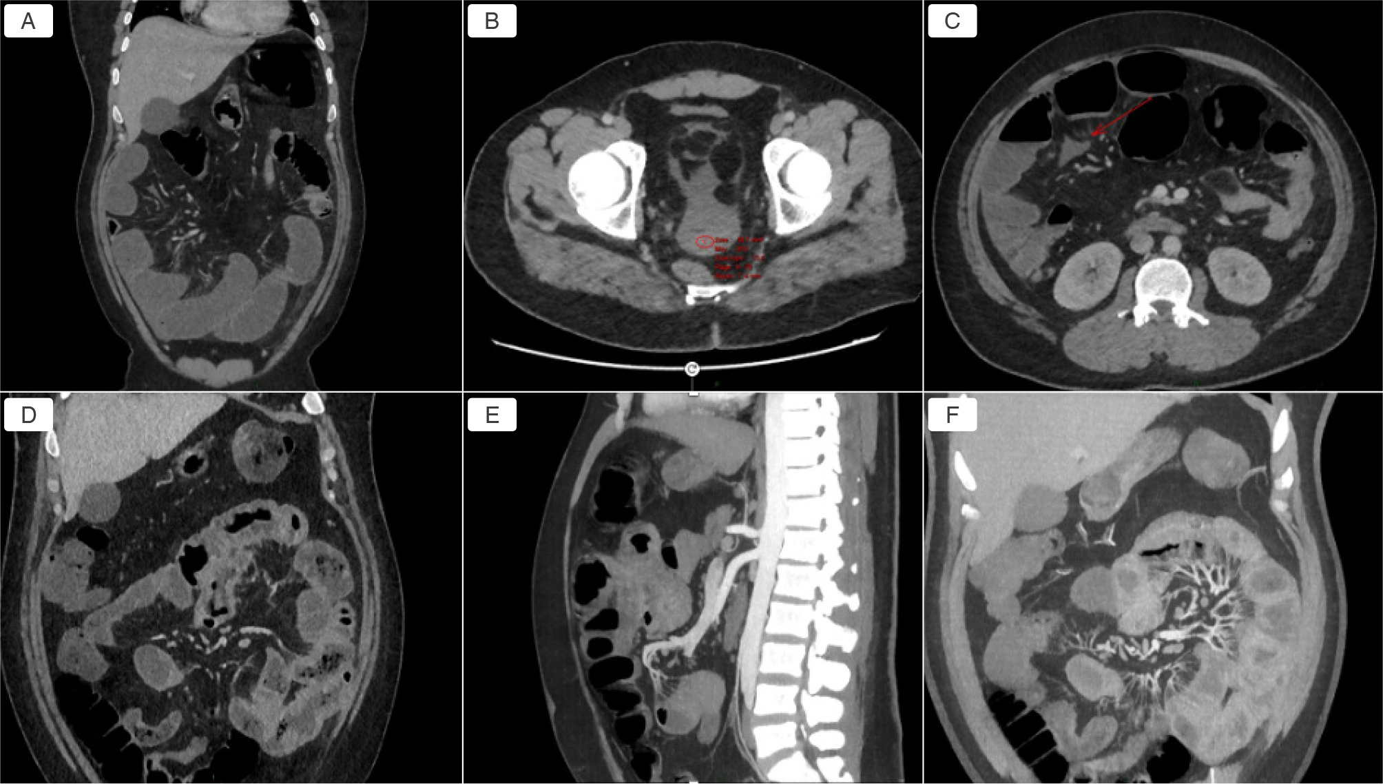
Abdominal computed tomography (CT) imaging (Fig. 1) demonstrated diffuse jejunal and ileal loop dilatation without an identifiable transition point, consistent with functional small-bowel obstruction. A small amount of spontaneously hyperdense pelvic free fluid (39 HU) was also observed, initially considered suggestive of a minor hemoperitoneum. However, there was no clinical evidence of active bleeding and hemoglobin levels remained stable throughout hospitalization. Associated findings included circumferential mural thickening with submucosal oedema and mesenteric venous hyperaemia, while mesenteric arterial vasculature remained patent and unremarkable.
(click to enlarge)
Figure 1. Figure A) Contrast-enhanced abdominal CT scan (portal venous phase, coronal view) showing dilatation of jejunal and ileal loops without a transition point, consistent with functional small-bowel obstruction; B) Contrast-enhanced abdominal CT scan (portal venous phase, axial view) showing a small amount of hyperdense free fluid in the pouch of Douglas (39 Hounsfield units); C) Contrast-enhance abdominal CT scan (portal venous phase, axial view) showing mesenteric fluid infiltration (red arrow); D) Contrast-enhanced abdominal CT scan (portal venous phase, coronal view) showing small-bowel wall thickening with areas of submucosal edema in the jejunum; E) Contrast-enhanced abdominal CT scan (portal venous phase, sagittal view with MIP reconstruction) showing normal morphology and patency of the superior and inferior mesenteric arteries; F) Contrast-enhanced abdominal CT scan (portal venous phase, coronal view with MIP reconstruction) showing normal morphology and patency of distal branches of the superior mesenteric artery, associated with mesenteric venous hyperemia.
Given the exclusion of infectious, neoplastic, and other inflammatory causes, a diagnosis of AOSD flare with intestinal involvement presenting as acute pseudo-obstruction was retained.
Empiric antibiotics were discontinued due to lack of efficacy. Immunosuppressive therapy was initiated with prednisone (1 mg/kg/day) and anakinra (100 mg/day subcutaneously). Clinical improvement was rapid, with resolution of fever within 48 hours, progressive normalization of bowel function, and disappearance of the rash. Inflammatory markers significantly decreased, with normalization of the C-reactive protein and reduction of ferritin levels to 1265 µg/l (glycosylated fraction 44%) at discharge. Corticosteroids were progressively tapered, and anakinra was continued with plans for spacing depending on clinical stability.
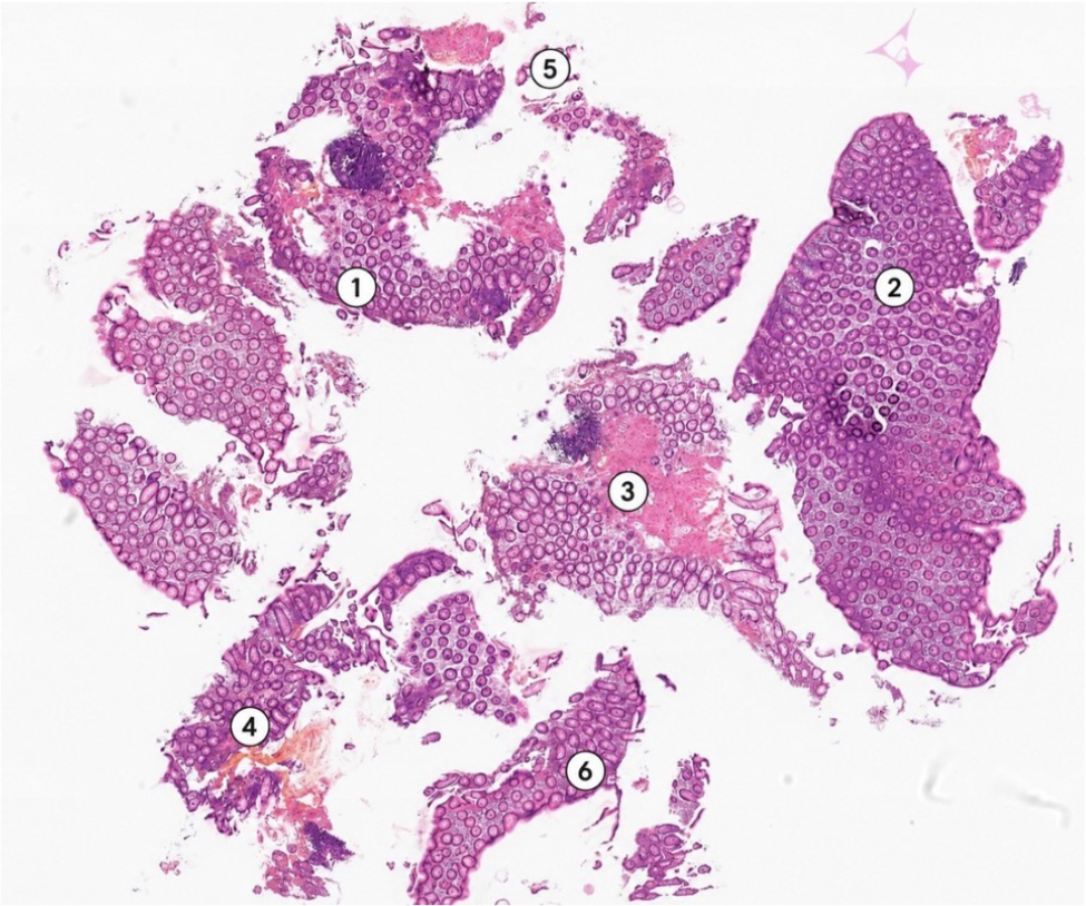
Entero-MRI performed on day 6 of treatment was unremarkable. Colonoscopy performed on day 10 was macroscopically normal, while histology revealed interstitial colitis with inflammatory infiltrates (Fig. 2).
(click to enlarge)
Figure 2. Microscopic examination of a colonic biopsy, hematoxylin–eosin stained.
This image demonstrates preserved crypt architecture with moderate inflammatory cell infiltrates in the lamina propria.
At the latest follow-up, more than one year after the initial episode, the patient remained in sustained clinical remission, with complete discontinuation of both corticosteroids and anakinra.
DISCUSSION
This case illustrates a rare but clinically significant manifestation of AOSD presenting as acute intestinal pseudo-obstruction. Gastrointestinal involvement in AOSD remains uncommon and poorly characterized, with only a limited number of cases reported in the literature, including presentations with duodenojejunal inflammation or functional bowel obstruction[2]. In another case reported by Hori et al., intestinal pseudo-obstruction was an inaugural manifestation of AOSD[3]. More severe presentations have also been described, including a fatal case associated with HLH and multiorgan failure[4]. In contrast, our patient had no significant organ failure and showed rapid improvement after corticosteroids and early IL-1 blockade. Finally, Manzotti et al. described chronic duodenojejunal inflammation[5], while our case was characterized by an acute obstructive presentation. These previous reports highlight the heterogeneous spectrum of gastrointestinal involvement in AOSD, ranging from chronic inflammation to acute pseudo-obstruction, either inaugural or associated with severe systemic manifestations such as HLH or coagulopathy. In contrast, our case involved acute pseudo-obstruction during a flare of established AOSD, with marked cytokine activation but without major associated organ failure.
In our patient, the diagnosis of AOSD flare was supported by marked systemic inflammation, with neutrophilic leukocytosis, hyperferritinaemia with low glycosylated fraction[2] after thorough exclusion of infectious, neoplastic, and autoimmune causes. Another key element in this case is the markedly elevated IL-18 level, which has emerged as a major biomarker of disease activity in AOSD[6]. Recent data from large cohorts have shown that IL-18 levels are significantly higher in AOSD compared to other autoinflammatory or autoimmune conditions, making it a useful diagnostic and discriminative marker[7].
Several differential diagnoses were carefully considered. Notably, cases of misdiagnosis between Crohn’s disease and AOSD have been reported[8]. In our patient, Crohn’s disease was ruled out despite potential overlapping inflammatory pathways involving IL-18, as imaging and endoscopic findings were not suggestive.
Beyond its diagnostic value, IL-18 plays a central role in the pathophysiology of AOSD by promoting macrophage activation and amplifying the cytokine cascade, particularly through interferon-γ pathways. Extremely high IL-18 levels have been associated with severe systemic inflammation and an increased risk of HLH[9].
In our patient, the very high IL-18 level may also support the hypothesis of cytokine-mediated intestinal dysfunction. Experimental and clinical data suggest that cytokine-driven inflammation, particularly involving IL-1 and TNF-α, may impair intestinal smooth muscle contractility and disrupt enteric nervous system function, leading to dysmotility[10]. Additionally, elevated IL-18 levels—frequently observed in active AOSD—may contribute to intestinal inflammation and mucosal immune activation[11], potentially explaining the association with interstitial colitis and elevated faecal calprotectin observed in our patient.
From a clinical perspective, this case highlights the importance of considering AOSD in cases of unexplained intestinal pseudo-obstruction associated with systemic inflammation. Early recognition is critical, as prompt initiation of targeted immunomodulatory therapy, particularly IL-1 blockade with anakinra, can lead to rapid clinical improvement and may prevent progression to severe complications such as HLH.