A Rare Case of Spinal Sarcoidosis Presenting as Multiple Bone Marrow Oedematous Lesions
  • Anat Bel-ange
    Department of Internal Medicine "C", Assaf Harofeh Medical Center, affiliated to Sackler Medical School, Tel Aviv University, Zerifin, Israel
  • Sigal Tal
    Department of Radiology, Assaf Harofeh Medical Center, affiliated to Sackler Medical School, Tel Aviv University, Zerifin, Israel
  • Micha Rapoport
    Department of Internal Medicine "C", Assaf Harofeh Medical Center, affiliated to Sackler Medical School, Tel Aviv University, Zerifin, Israel

Keywords

Sarcoidosis, sarcoid bone lesions, spine, MRI, bone marrow oedema

Abstract

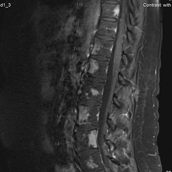
Sarcoidosis is a systemic disorder that most commonly affects the lungs. Bone involvement is rare, and spinal involvement is even more rare. The presence of focal lesions of the vertebrae is highly suspicious of advanced malignancy. However, malignant metastatic spread to the spine involves the vertebral cortex rather than the bone marrow itself, a distinction that is often missed and therefore misleading. We describe here a middle-aged woman with multiple focal oedematous lesions of the bone marrow suspected of being advanced malignancy but finally diagnosed as a rare case of spinal sarcoidosis.

VIEW THE ENTIRE ARTICLE

References

  • Eriksen EF. Treatment of bone marrow lesions (bone marrow edema). BoneKEy Rep 2015;4:755.

  • Bauerle T, Semmler W. Imaging response to systemic therapy for bone metastases. Eur Radiol 2009;19:2495–2507.

  • O'Sullivan GJ, Carty FL, Cronin CG. Imaging of bone metastasis: an update. World J Radiol 2015;7:202–211.

  • Hanrahan CJ, Shah LM. MRI of spinal bone marrow: part 2, T1-weighted imaging-based differential diagnosis. AJR Am J Roentgenol 2011;197:1309–1321.

  • Valencia MP, Deaver PM, Mammarappallil MC. Sarcoidosis of the thoracic and lumbar vertebrae, mimicking metastasis or multifocal osteomyelitis by MRI: case report. Clin Imaging 2009;33:478–481.

  • Caobelli F, Gabanelli SV, Brucato A, Pizzocaro C, Soffientini A, Giubbini R, et al. Unsuspected active sarcoidosis diagnosed by 18F-FDG PET/CT during the search for a primary tumour in a patient with bone lesions. Nucl Med Mol Imaging 2013;47:205–207.

  • Lefere M, Larbi A, Malghem J, Vande Berg B, Dallaudiere B. Vertebral sarcoidosis: long-term follow-up with MRI. Skeletal Radiol 2014;43:1185–1190.

  • Rua-Figueroa I, Gantes MA, Erausquin C, Mhaidli H, Montesdeoca A. Vertebral sarcoidosis: clinical and imaging findings. Semin Arthritis Rheum 2002;31:346–352.

  • Rodman T, Funderburk EE, Jr., Myerson RM. Sarcoidosis with vertebral involvement. Ann Int Med 1959;50:213–218.

  • Johnson AK, Johnson JM, Ames E, Filippi C. Spontaneous clinical and radiological resolution of vertebral sarcoidosis: a case report. Spine 2012;37:E414–416.

  • Mehrotra P, Muller M, Higgins B, Zammit-Maempel I. An unusual cause of widespread lytic bone lesions caused by sarcoidosis. J Radiol Case Rep 2011;5:1–7.

  • Soussan M, Augier A, Brillet PY, Weinmann P, Valeyre D. Functional imaging in extrapulmonary sarcoidosis: FDG-PET/CT and MR features. Clin Nucl Med 2014;39:e146–159.

  • Mostard RL, Prompers L, Weijers RE, van Kroonenburgh MJ, Wijnen PA, Geusens PP, et al. F-18 FDG PET/CT for detecting bone and bone marrow involvement in sarcoidosis patients. Clin Nucl Med2012;37:21–25.
  • Views: 1192
    HTML downloads: 729
    PDF downloads: 463


    Published: 2018-06-21
    Issue: Vol 5 No 8 (view)


    How to cite:
    1.
    Bel-ange A, Tal S, Rapoport M. A Rare Case of Spinal Sarcoidosis Presenting as Multiple Bone Marrow Oedematous Lesions. EJCRIM 2018;5 doi:10.12890/2018_000907.

    Similar Articles

    You may also start an advanced similarity search for this article.